The growing use of smart technologies has revolutionized the world and how people live in it, making people’s lives easier, faster, and more convenient. Advanced technologies, artificial intelligence (AI), machine learning, big data analytics and the Internet of things (IoT) have paved the way for the development of sophisticated applications designed for specific environments such as manufacturing, logistics and planning, as well as business operations. They are used to improve manufacturing processes, provide more sophisticated data analytics, automate manual tasks and improve quality control. Known as the Fourth Industrial Revolution (4IR) era, these technologies and devices are widely applied for innovation and value creation across many industries.[1]
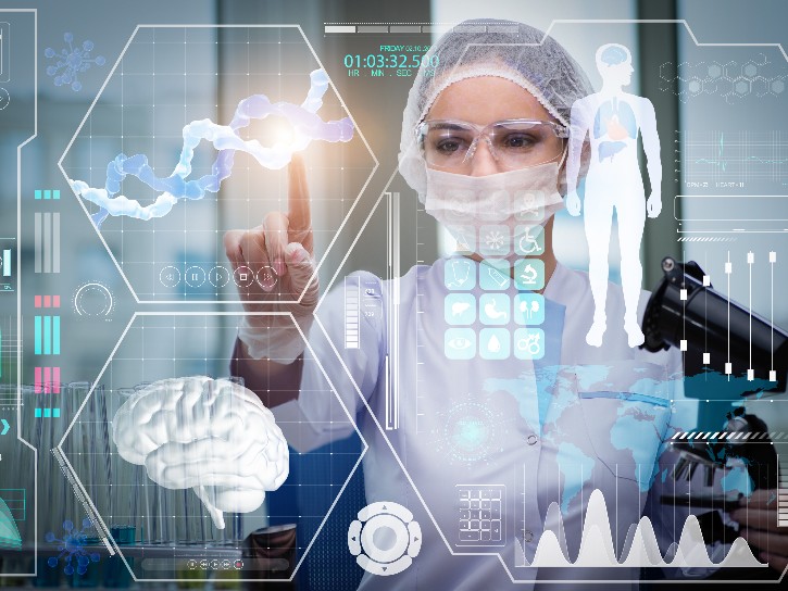
The healthcare industry has not been left behind. Widespread applications of advanced technologies and AI in healthcare institutions are supporting efforts to improve care, service quality, and the efficiency of medical resources and staff. The adoption of some of these technologies in radiology has not only been effective in helping radiologists process large volumes of imaging data with advanced image reconstruction algorithms, but also holds potential for improving the consistency and accuracy of patient imaging, diagnoses, alleviating some of the work-related stress and strain on radiology staff, and improving the overall patient experience.
Industry leaders in imaging, partners in computing technology, and independent start-ups alike are working together to bring innovation to healthcare using advanced digital technologies and AI to create value and improve efficiencies.
Health, Life Sciences and Emerging Technologies
Specifically in radiology, innovative technologies and AI are being used to help clinicians triage critical cases in X-ray, improve consistency, and eliminate scanning inefficiencies in magnetic resonance imaging (MRI), and evolve advanced visualization and image processing capabilities with cloud-based, enterprise level solutions that support today’s radiology needs.
Building AI applications for use at the point of care
The importance of quickly triaging patients toward the right treatment path can be critical to a patient’s outcome. Imaging plays a vital role in determining that path; however, with limited resources to read images, or high volumes of patients waiting for diagnoses, time to diagnosis can be delayed.
These tools can help automate some of the technologist’s manual tasks and help provide reading physicians with clinical decision support.
A new AI tool embedded on mobile X-ray systems supports the exam workflow, and helps clinicians prioritize critical cases. During the image acquisition, the AI automatically orients each image properly, so the technologist does not have to do that extra work before sending the image to the PACS, which is a significant time savings. The AI algorithm also quickly reviews the image taken against the imaging protocol that was ordered to ensure the correct image was taken, and calls attention to any image imperfections, such as a clipped lung, which is a common image error. The technologist is able to correct these errors while the patient is still in the room. These solutions, though minor, save a great deal of time for the technologist, and help reduce the need for retakes by getting the right image for each patient at the point of care.
Because X-ray exams are frequently ordered as urgent, or STAT, even a small delay in diagnosis and treatment can have a profound impact on patient outcomes. To help improve speed to diagnosis, this AI solution helps draw a finer separation between STAT and critical patients to allow timely reporting and more accurate patient management. It works by detecting subtle or complex patterns within patient images at the point of care, flags suspicious findings, and prioritizes those cases for radiologist review, to help with improvements across the board in efficiency, quality, and clinical accuracy. With no additional delay or processing time, the AI algorithm provides triage notifications to the radiologist that arrive in PACS at the exact same time as the DICOM image.
Using AI to automate workflow, and improve consistency and accuracy in neuro MR imaging
Neuroimaging or brain imaging with MRI is used to image the structure, function, or pharmacology of the nervous system. Structural imaging is often used to diagnose intracranial diseases, such as brain tumors, while functional imaging is used to diagnose metabolic diseases. To monitor these diseases over time, multiple MRI scans are often needed. Before an MRI technologist can scan a patient, they have to manually specify the slices they want the MRI to acquire. This process can take several minutes of tweaking and adjusting, leaving a patient waiting anxiously in the MRI scanner and adding unnecessary steps to set up each scan. It can also introduce inconsistencies into images taken over time if parameters or positioning are slightly different each time a patient gets scanned, making it challenging to accurately monitor disease progression or treatment.
For the imaging studies to be evaluated longitudinally, they must image the same area of interest each time. If parameters or positioning are slightly different each time a patient gets scanned, it can be challenging to accurately monitor disease progression or treatment. The processes of result validation and reproducibility of different neuroimaging analyses and statistical maps in MRI are often difficult because of a number of factors, some of which include changes to the patient between scans, presence of noise in the imaging data, and variations in study designs, sample sizes and sampling protocols.[2]
An AI-based, automated workflow tool is now available that uses state-of-the-art AI to precisely identify and align MR scans for diagnostic neuroimaging. It automates the area of interest and reduces redundant, manual steps that are typically completed by the MRI technologist. Streamlining the MRI workflow, the AI technology enables consistent, repeatable scan alignment to help physicians better monitor a patient across longitudinal studies which may be several months apart. It also reduces the set-up time for MRI studies and helps produce images that have less variability between technologists and between scans. This lowers the chances a patient will be recalled due to incorrect slice placement.
Developing scalable software solutions for flexible work environments in radiology
Advanced technologies are not only changing the way radiologists work, but also where they can work. These new technologies are now enabling image processing software, typically only available on the local workstation that was installed with any given imaging system, to be used on any workstation or pc, onsite or virtually. Employed this way, AI-based reconstruction algorithms for all modalities can be accessed through advanced visualization and image processing software solutions that are server- or cloud-based, and able to be used enterprise-wide. A single user can work with the images from multiple modalities or workstations, and the number of simultaneous users can be scaled to the meet needs of the department.
The benefits of using server- or cloud-based visualization software solutions include improved utilization awareness and improved resource utilization, both of human resources as well as equipment within the radiology department. The ease and flexibility of using the software from any system also supports virtual work environments and improved collaboration between clinicians, enabling a more efficient workflow, potentially easing work-related stress and strain on radiologists, and potentially improving patient outcomes.
Increasing sustainable efficiencies with advanced technologies and AI
The continued development and use of rapidly advancing technology and AI tools enable radiology to double-down on efforts to increase efficiency, improve workflow and create better health outcomes for patients. Leveraging advanced technologies and AI to provide clinical decision support across clinical applications could further improve the accuracy and consistency in medical imaging and continue to positively impact patient outcomes.
For more information on Critical Care Suite, GE Healthcare’s AI-enabled X-ray tool, click here.
For more information on AIR x™, GE Healthcare’s MR workflow Automation tool, click here.
For more information on AW, GE Healthcare’s Advanced Visualization Solution, click here.

GE Healthcare Experience is live! Your one-stop source for all information about GE Healthcare at RSNA 2021… from news, webinars, live broadcasts and more than 60 new products. Get your GE Digital Pass now! https://gehealthcare.smh.re/0K7
REFERENCES
[1] Lee D, Yoon SN. Application of Artificial Intelligence-Based Technologies in the Healthcare Industry: Opportunities and Challenges. Int J Environ Res Public Health. 2021;18(1):271. Published 2021 Jan 1. doi:10.3390/ijerph18010271
[2] Dinov ID. Neurological imaging: statistics behind the pictures. Imaging Med. 2011;3(4):423-432. doi:10.2217/iim.11.37