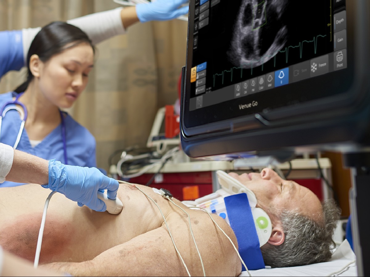
Until the 1990s, critically ill patients who needed rapid disease assessment didn't have many options. They were limited to invasive modalities and traditional radiographic imaging—both of which pose risks to the patient. Point of Care Ultrasound (POCUS) changed that.
What is POCUS's role in the modern medical landscape? Here's how this technology has transformed not only emergency medicine but also the practice of all medicine.
Evaluating Patients Before POCUS
Prior to the availability of POCUS, practitioners relied on other methods to assess critically ill patients—for instance, central lines for measuring central venous pressure in hypotensive patients, Swan-Ganz catheters for gauging cardiac pressures, and diagnostic peritoneal lavage for diagnosing hemoperitoneum in trauma patients. Although these modalities are useful, they are typically neither time sensitive nor straightforward to interpret, which can negatively affect outcomes for patients with elevated risks.
Traditional radiographic imaging such as the CT scan or MRI, both of which are sensitive and specific for diagnosis, can be time consuming and physically demanding. They require moving a patient to radiology, performing the scan, and then waiting for the radiologist to read and post the results. This method is often helpful for stable patients, but critically ill and unstable patients cannot always safely move to radiology for care.
A History of Ultrasound
Ultrasound was discovered in the 1790s as Italian physiologist Lazzaro Spallanzani studied sonar in bats.2 A growing understanding of the principles behind ultrasound waves helped set a foundation for their eventual use in medicine—in the 1940s, neurologist Karl Dussik used ultrasound in an attempt to detect brain tumors.3 The technology began to find its place in the clinical setting in the 1960s. Nevertheless, there were many barriers for the use of ultrasound, including: 4
The size of the devices. They were large and bulky.
Patients had to be submerged in water during exams. Ultrasound waves move much faster in water than through air; water provides clearer, more stable images.
Difficulty reading images. Ultrasound images were often poor quality, as they were still images of a moving object.
All of these challenges made ultrasounds useful only as a traditional imaging modality performed in the radiology suite. During the 1970s, ultrasound technology was refined with the development of better transducers and improvements in image quality. These developments created opportunities for uses in more clinical settings, including in obstetrics, cardiology, and radiology. However, delays continued between image acquisition and interpretation, limiting it as a point-of-care modality.
By the 1980s, ultrasound technology had improved enough to allow for real-time scanning and image display, making its use more feasible in point of care. However, the 1990s brought dramatic improvements in size, portability, and technology, allowing for ultrasound to be practical for use at the bedside.6
Since then, POCUS has been instrumental in emergency medicine, trauma surgery, and critical care. Specialists recognized that a portable ultrasound probe allowed them to rapidly assess critically ill patients at the bedside. When the FAST exam was incorporated into advanced trauma life support (ATLS), POCUS was recognized as a standard of care.
Focused Assessment with Sonography in Trauma
One example of POCUS's advantages in trauma care is the Focused Assessment with Sonography in Trauma (FAST) exam, which replaces the invasive diagnostic peritoneal lavage (DPL) for determination of hemoperitinuem in trauma patients.1 Hemoperitoneum can be a surgical emergency, depending on the source of the bleeding. With DPL there is a high sensitivity for detecting hemoperitoneum but a low specificity if the bleeding requires the patient to go to the operating room. Since the methodology shows that there is bleeding but not the location, beside ultrasound can help avoid an exploratory laparotomy.
Beyond trauma patients, POCUS has changed the way physicians approach critically ill patients more broadly:
It can illustrate life-threatening conditions, helping to improve patient care, avoid invasive procedures, and decrease costs.
Exams are focused to answer a specific question, allowing for shorter patient evaluations.
It is quick to perform, taking only a few minutes to complete an exam.
Exams are characterized by one or two clearly recognized findings, making for easier and faster clinical decision-making.
The technology is easy to learn.
It can directly alert clinicians to whether the patient needs fluids or pressors.
It is small and portable, allowing the exam to travel to the patient.
Additional Emergency Uses
The FAST exam was only the first standardized use of emergency POCUS. It quickly took off in other areas of emergency medicine assessment as well. Many surgical emergencies originate in the thorax, abdomen, and pelvis—all areas which are relatively easy offshoots of the FAST exam.
Diagnosis of Ruptured Abdominal Aortic Aneurysm
Rupture of abdominal aortic aneurysms (AAA) is a true surgical emergency with a very high mortality rate, reaching 75% of those patients who do make it to the operating room.5 Ruptured AAAs can cause catastrophic intraperitoneal bleeding.
These patients are typically unstable, and getting them to the operating room quickly is key. Patients with suspected AAA require rapid evaluation, allowing little time for transfer to radiology for CT scans. POCUS is an effective modality for diagnosis. Systematic reviews of POCUS for AAA show sensitivities of 94% to 100% and specificities of 98% to 100%, which are similar to screening ultrasounds performed in radiology suites.5
Diagnosis of Ectopic Pregnancy
Ectopic pregnancy is an obstetric emergency that can cause massive bleeding and require immediate surgery. The use of POCUS over traditional ultrasound has been shown to decrease emergency room time and time to surgery for patients. Whereas the average emergency room treatment time in the POCUS group was 157.9 minutes to the radiology group of 206.3 min., and the median time to the operating room for ruptured ectopic pregnancies was 203.0 min vs 293.0 min.7
Advancement of POCUS Outside the Emergency Department
Early adopters and leaders in POCUS extend to musculoskeletal, anesthesiology, and critical care. These teams have used POCUS for resuscitation, procedural guidance, evaluation of new signs and symptoms, and for therapy and monitoring.
Resuscitation
POCUS can be used for resuscitation both during cardiac arrest and as a guideline for fluid administration in hemodynamically unstable patients. In these settings, POCUS can be used to assess why a patient is unstable—either related to preload, cardiac issues, or afterload reasons. This allows physicians to more rapidly treat and stabilize them, in turn decreasing the risk of mortality.
Procedural Guidance
Anesthesiologists and critical care physicians use the placement of invasive lines to more fully monitor critically ill patients. This includes procedures such as setting central venous lines, arterial lines, and dialysis catheters. Prior to the wider use of POCUS, physicians used anatomical landmarks for these placements. Now, ultrasound's ability to directly visualize the anatomy has decreased procedure times as well as complication rates.
Evaluation
Patients with chest pain and dyspnea frequently enter emergency rooms, intensive care units, and post-operative care units. There is a long differential diagnosis for these cases involving myocardial infarction, pneumothorax, pericardial effusion, pleural effusion, pulmonary embolism, and pain. POCUS can help differentiate between these and guide treatment more quickly than can lab findings and traditional imaging modalities. A quick scan with POCUS allows a physician to search for pericardial fluid, discrepancies in right and left ventricula size, cardiac dysfunction, pleural sliding, and aortic root dilation. Positive findings in any of these areas can point the physician toward the correct diagnosis.8
Therapeutic or Monitoring Indications
All patients receive noninvasive monitors including those for blood pressure, pulse oximetry, and heart rate. Critically ill patients often require invasive monitoring such as arterial lines, central venous lines, and Swan-Ganz catheters. POCUS's guidance can help clinicians place the lines more quickly and with fewer complications.
Beyond Critical Care
POCUS is now gaining traction in other areas of medicine for more effective care and clinical decision-making:
Anesthesiologists use POCUS for placement of nerve blocks for pain control perioperatively. In the past, these blocks were placed using anatomic landmarks and nerve stimulation, but POCUS has improved block success and decreased complication rate.
Gastroenterologists use POCUS to monitor obstruction within the biliary tract and evaluate for biliary disease.
Nephrologists may use POCUS for many reasons, including to assess the placement of peritoneal dialysis catheters or evaluate for cysts in the kidney or hydronephrosis.
Abdominal pain is a common patient complaint. POCUS can help clinicians evaluate for many concerns, including ectopic pregnancy, appendicitis, and intraperitoneal infection.
Obstetrics and gynecology may turn to POCUS for evaluation of fetal position prior to and during labor.
Physical medicine and rehabilitation physicians as well as sports medicine physicians often use POCUS to guide joint injections.
Ultrasound is the primary method of diagnosing deep vein thrombosis. Now, POCUS has moved that to the bedside.
What is POCUS's ultimate place in medical history? Although bedside ultrasound took 50 years to come to fruition, its accessibility and cost have allowed it to become a versatile diagnostic modality. POCUS continues to offer advantages to physicians. Because of its ease of use and short time to master, every physician has the ability to bring POCUS to their practice.
Sources:
- Bloom BA, Gibbons RC. Focused assessment with sonography for trauma. [Updated 2021 Jul 31]. StatPearls. Treasure Island (FL): StatPearls Publishing; Jan 2022. https://www.ncbi.nlm.nih.gov/books/NBK470479/.
- CME Science. Who Invented Ultrasound? CME Science. https://cmescience.com/who-invented-ultrasound/. Accessed Apr 20, 2022.
- Evolution of Point of Care Ultrasound. Radiology Key. https://radiologykey.com/evolution-of-point-of-care-ultrasound/. Accessed April 18, 2022.
- Kaproth-Joslin K, Nicola R, Vikram S. The history of US: From bats and boats to the bedside and beyond: RSNA Centennial Article. Radiographics. Mar 2015; 35(3):657-679. https://pubs.rsna.org/doi/full/10.1148/rg.2015140300.
- Jeanmonod D, Yelamanchili VS, Jeanmonod R. Abdominal Aortic Aneurysm Rupture. [Updated Aug 2021]. StatPearls. Treasure Island (FL): StatPearls Publishing; Jan 2022. https://www.ncbi.nlm.nih.gov/books/NBK459176/.
- John K, Hoffenberg S, Smith S. History of emergency and critical care ultrasound: The evolution of a new imaging paradigm. Critical Care Medicine. May 2007;35(5):126-130. https://pubmed.ncbi.nlm.nih.gov/17446770/.
- Stone BS, Muruganandan KM, Tonelli MM. Impact of point-of-care ultrasound on treatment time for ectopic pregnancy.The American Journal of Emergency Medicine. Nov 2021;49:226-232 https://www.sciencedirect.com/science/article/abs/pii/S0735675721004551
- Melgarejo S, Noble V, Schaub, A, Point of care ultrasound: An overview. American College of Cardiology. Oct 2017. https://www.acc.org/latest-in-cardiology/articles/2017/10/31/09/57/point-of-care-ultrasound. Accessed Apr 20, 2022.

