Blood clots, including deep vein thrombosis (DVT) and pulmonary embolism, represent a dangerous and common complication for many hospitalized patients.
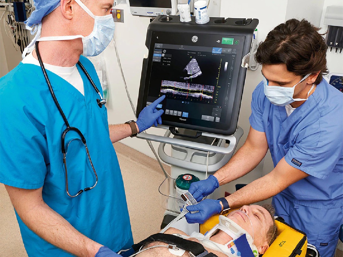
Increasingly, emergency departments (EDs) are turning to Point of Care Ultrasound (POCUS) to improve patient outcomes. Using ultrasound to find blood clots gives doctors another tool that they can bring directly to the bedside as they search for timely answers to help triage care.
Addressing the Risk of Blood Clots
The Centers for Disease Control and Prevention (CDC) estimate that about 900,000 people experience a blood clot every year—and about 100,000 people die from one.1 Early diagnosis of blood clots is critical to prevent death.2
DVT starts in a deep vein in the leg or arm. Part of the clot can break off and travel to the lungs, causing a pulmonary embolism, which can be fatal without treatment. DVT can also lead to long-term complications, making detecting and managing clots critical to patient outcomes.
Anyone can be at risk of developing a blood clot, but certain patients in the ED may have a higher risk—and some may already have a blood clot without knowing it. Medical conditions, recent surgery, and certain treatments affect the body's blood flow and clotting ability, which leads to more risk of blood clots. Pregnant patients, people with cancer, patients who are sedentary or immobilized, and patients who have recently had surgery are at higher risk and may be candidates for DVT monitoring. People who have had a blood clot in the past are also at risk of developing another one.2
About half of people with DVT have no symptoms, and they can develop a pulmonary embolism without any noticeable signs. That's why pulmonary embolism is often referred to as a silent killer. The lack of symptoms makes detecting DVT even more crucial. A blood clot in a deep vein doesn't cause a stroke. However, as many as half of the people who develop DVT experience long-term effects. Called post-thrombotic syndrome, the lasting impact of the condition can be debilitating.2
Finding Timely Answers with POCUS
Duplex ultrasonography, often performed by a sonographer and interpreted by radiology, sits alongside contrast venography as the standard diagnostic tools for DVT. Yet time is of the essence in a bustling ED, and care teams may need faster ways to triage patients.
Using bedside ultrasound helps clinicians find blood clots with accuracy comparable to that of duplex ultrasonography—and do it quickly. Findings from multiple studies show:
The average time from request to completion of the ultrasound study was 15 minutes in a busy ED.4
The time from triage to a patient being discharged from the ED or admitted to the hospital was 95 minutes when an emergency physician performed the ultrasound compared with 220 minutes when radiology performed the study.4
The median time from order to completion of POCUS was 5.8 hours compared with 11.5 hours from the time of order to finalized radiology report.5
Not all hospitals have access to radiologists or lab studies 24 hours a day. Patients may need to be hospitalized or receive unnecessary anticoagulation based on a suspected DVT until the possibility is ruled out or definitively diagnosed. POCUS is readily available in many hospitals; without access to radiology, this tool can get care teams the answers they need to move patients through the ED.3
Learning Lessons from POCUS during COVID-19
The COVID-19 pandemic cemented a need to move patients through the ED. In response, POCUS highlighted the ability to do just that. People with COVID, particularly those requiring hospitalization, are at increased risk of thrombotic events such as DVT. This made screening crucial. As patients overwhelmed EDs, hospitals relied on bedside ultrasound to find blood clots.
Beyond being reproducible and portable, ultrasound can help lower the risk of infection by keeping care at the bedside rather than forcing care teams to transfer patients. The sensitivity of bedside ultrasound when diagnosing lower extremity DVT varied from 84% to 97% with a specificity greater than 95%.6
The effectiveness of POCUS to screen for DVT during the pandemic demonstrated the versatility of this tool in bringing diagnostic imaging to patients, help clinicans get a faster answer, and moving patients into appropriate care without having to wait for lab results. Those lessons can apply to future disease outbreaks, and other emergencies that strain hospital EDs.
Transforming Care with Ultrasound for DVT
A POCUS exam to look for DVT involves compressing the common femoral vein (CFV) and the popliteal vein (PV) or compressing the CFV, PV, and the superficial femoral vein. When a blood clot is present, the vein doesn't fully compress.4
Some patients, such as those at high risk for DVT, may need screening. Others may show DVT symptoms that need analysis. The location of the clot—whether above the knee, below the knee, or in the venous system—allows clinicians to direct patients to the appropriate therapy.7
Pulmonary embolism is one of the biggest risks associated with DVT. However, POCUS in the ED may be able to exclude centrally located pulmonary embolism.8 A proximal positive result with lower limb POCUS has a high positive predictive value for pulmonary embolism.9 Additionally, it can be performed on patients who aren't able to undergo computed tomographic pulmonary angiography for embolism diagnosis. Plus, identifying DVT can help patients receive early anticoagulation therapy to reduce the risk of pulmonary embolism.8,9
With the help of ultrasound, physicians can also distinguish conditions that can look similar to DVT. When DVT symptoms do occur, they can include pain, swelling, redness, and warmth at the clot. These symptoms often appear similar to cellulitis, vasculitis, and acute arterial occlusion, but ultrasound evaluation helps rule out these conditions.2
A rouleaux formation may also appear on ultrasound. Created when red blood cells stack on top of each other, it lies over venous valves, looking like a spontaneously echogenic blood flow. Rouleaux formations are common and typically not of clinical concern—but they can signal a proximal DVT. The presence of this formation is not enough to diagnose DVT, so further examination is required to rule out DVT. Veins with a rouleaux formation can be compressed, which is not the case when a blood clot is present.4,10
The accuracy, timeliness, and portability of POCUS makes it an essential tool for physicians in the ED as part of their assessment for DVT. Identifying DVT early can help patients access appropriate treatment quickly, improve ED throughput, and help prevent complications.
In the chaos of emergency departments, having a system that is ready to go when you need it is imperative. https://venue-pocus.gehealthcare.com/emergency-ultrasound
References
1. Centers for Disease Control and Prevention. Impact of blood clots on the United States. CDC.gov. https://www.cdc.gov/ncbddd/dvt/infographic-impact.html. Accessed May 17, 2022.
2. Centers for Disease Control and Prevention. What is venous thromboembolism? CDC.gov. https://www.cdc.gov/ncbddd/dvt/facts.html. Accessed May 17, 2022.
3. Centers for Disease Control and Prevention. Diagnosis and Treatment of Venous Thromboembolism https://www.cdc.gov/ncbddd/dvt/diagnosis-treatment.html. June 9, 2022
4. Varrias D, Palaiodimos L, Balasubramanian P, et al. The use of point-of-care ultrasound (POCUS) in the diagnosis of deep vein thrombosis. Journal of Clinical Medicine. 2021;10(17):3903. https://www.ncbi.nlm.nih.gov/pmc/articles/PMC8432124/.
5. Fischer EA, Kinnear B, Sall D, et al. Hospitalist-operated compression ultrasonography: a point-of-care ultrasound study (HOCUS-POCUS). Journal of General Internal Medicine. 2019;34:2062–2067. https://link.springer.com/article/10.1007/s11606-019-05120-5.
6. Kapoor S, Chand S, Dieiev V, et al. Thromboembolic events and role of point of care ultrasound in hospitalized covid-19 patients needing intensive care unit admission. Journal of Intensive Care Medicine. 2021;36(12):1483-1490. https://journals.sagepub.com/doi/10.1177/0885066620964392.
7. Baker M, Anjum F, dela Cruz J. Deep venous thrombosis ultrasound evaluation. StatPearls. 2021. https://www.ncbi.nlm.nih.gov/books/NBK470453/.
8. Dwyer KH, Rempell JS, Stone MB. Diagnosing centrally located pulmonary embolisms in the emergency department using point-of-care ultrasound. American Journal of Emergency Medicine. 2018;36(7):1145-1150. doi:10.1016/j.ajem.2017.11.033.
9. Squizzato A, Galli L, Gerdes VE. Point-of-care ultrasound in the diagnosis of pulmonary embolism. Critical Ultrasound Journal. 2015;7:7. doi:10.1186/s13089-015-0025-5.
10. Blanco P, Volpicelli G. Common pitfalls in point-of-care ultrasound: a practical guide for emergency and critical care physicians. Critical Ultrasound Journal. 2016;8(1):15. doi:10.1186/s13089-016-0052-x.

