Today's care providers need to be able to accurately track uterine activity on a fetal monitor during labor. When mothers are induced, have a high BMI, or have certain complications, such as intrauterine growth restriction or pre-eclampsia, precise uterine monitoring is especially imperative. Still, even during normal labor with few or no complications, nurses and physicians alike want uterine activity tracings that are as accurate as possible.
For example, precise UA tracking is necessary when analyzing fetal heart tones, particularly if there are decelerations in the fetal heart rate. Additionally, a study in the Journal of Midwifery & Women's Health found that even though labor dystocia is the leading cause of U.S. cesarean births, it has been difficult for care providers to measure its development during labor.1 The study analyzed how UA monitoring can help nurses make more accurate predictions and gain a better understanding of how the condition develops, leading to improved outcomes for both mother and baby.
However, current monitoring solutions for tracking uterine activity on a fetal monitor do present a range of challenges—and this can lead to a disappointing and frustrating labor experience for everyone involved. As a labor and delivery nurse, you and your patients may face the following issues, and here is how a wireless monitoring option can help improve outcomes.
Monitoring Challenges to Patient Safety and Comfort During Labor
First and foremost, your priority is ensuring the safety of both mother and baby, which includes providing a comfortable experience, while getting the information you need to do your job effectively. When it comes to accuracy, the intrauterine pressure catheter is often considered the gold standard, as it is known to provide the most precise readings. It provides reliable data about ongoing contractions, their pressure, and frequency, and it can be the first to detect a possible problem for mother or baby. Still, this method of tracking UA requires membrane rupturing, which is invasive, often uncomfortable, and may not be feasible for all patients. It can also lead to complications, such as maternal fever, according to research from the American Journal of Obstetrics & Gynecology (AJOG).2
That said, even external monitoring technology comes with its own challenges. Take, for instance, placing a tocodynanometer (TOCO). This type of UA tracking tool often needs to be attached via wires to a fetal monitor and requires a tight belt around the patient's stomach. In addition to causing patient discomfort, the belt and monitor can then become dislodged when your patient moves around in bed, leaving you to frequently readjust the belt to ensure continuous and precise tracking. A Reproductive Sciences study found that patients with a high BMI might experience trouble with TOCO monitoring, as the tight fit might be simply unbearable, but more importantly, the device's efficacy diminishes with increased maternal BMI.3
The wired TOCO limits how much your patient can walk around while in labor, and ambulation has been shown to provide a range of benefits for patients in labor. A meta-analysis published by The Cochrane Pregnancy and Childbirth Group compared upright positioning (walking, sitting, standing, kneeling) with lateral or supine positions during the first stage of labor. It found that upright positions shortened the duration of the first stage of labor by approximately 1 hour and 22 minutes. Research from the American College of Obstetricians and Gynecologists also found that water immersion can lower pain scores for certain patients.4 During the first stage of labor, patients can consider bathing in a shower or tub without harm to themselves or baby. However, some monitoring devices can't be used in water.
Overall, being tethered to a monitor adds an extra layer of complexity that can significantly reduce the quality of a patient's labor.
Improving L&D Outcomes with Precise, Wireless Monitoring
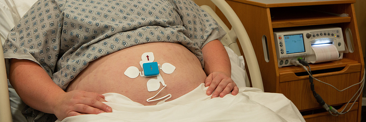
Uterine electromyography (EMG) signals, which report the electric activity of the uterine muscle, can help care providers find the right balance between obtaining accurate UA readings and making their patients comfortable. Additional AJOG research found that between the two noninvasive methods, EMG produced more precise and higher-quality UA tracings when compared to TOCO.5 In a separate AJOG study, patients reported higher satisfaction with this tracing method and it was found to be more effective on those with higher BMIs.6 Transmitted through wireless, adhesive surface electrodes placed on the patient's abdomen, EMG provides the accuracy and ease that care providers need, along with a range of additional benefits:
- EMG provides an alternative, less invasive method of UA monitoring. As obesity, and particularly morbid obesity, becomes more prevalent, external monitoring of contractions may not be possible. For these patients, EMG can provide an alternative, less invasive method of UA monitoring not affected by adipose tissue.
-
EMG empowers patients to create their ideal labor and delivery experience through freedom of movement. By enabling them to choose their laboring position or move around in bed as needed without tangling cords, you give patients the ability to make the right choices for themselves.
- EMG allows for optimal ambulation with a Bluetooth connection. Whether this means walking, swaying, or dancing around the labor and delivery room or hospital hallways, bathing in a shower or tub, or using the bathroom without assistance, patients can be as mobile as they want while being continuously monitored.
-
EMG improves the effectiveness of patient care. As a care provider, you have the freedom to help and support your patients without needing to constantly readjust or move wires or monitors. These devices can also simultaneously function to provide electronic fetal monitoring.
With the advent of new technologies to track uterine activity on a fetal monitor, patients are more empowered than ever before in their labor and delivery experiences. At the same time, you are able to optimize your workflows to ensure patient safety remains the priority. This not only leads to an improved birthing experience for the mother, but it also offers higher caliber surveillance for nurses and physicians to provide quality care.
References:
- Kissler KJ, Lowe NK, Hernandez TL. An integrated review of uterine activity monitoring for evaluating labor dystocia. Journal of Midwifery & Women's Health. June 2020;65(3):323-334. doi:10.1111/jmwh.13119
- Harper LM, Shanks AL, Tuuli MG, et al. The risks and benefits of internal monitors in laboring patients. American Journal of Obstetrics and Gynecology. July 2013;209(1):38.e1-38.e6. doi:10.1016/j.ajog.2013.04.001
- Aina-Mumuney A, Hwang K, Sunwoo N, et al. The impact of maternal body mass index and gestational age on the detection of uterine contractions by tocodynamometry. Reproductive Sciences. December 2015;23(5):638-643. doi:10.1177/1933719115611754
- Bryant AS, Borders AE. Approaches to limit intervention during labor and birth. The American College of Obstetricians and Gynecologists Clinical Committee Opinion. February 2019;133(2). https://www.acog.org/clinical/clinical-guidance/committee-opinion/articles/2019/02/approaches-to-limit-intervention-during-labor-and-birth
- Euliano TY, Nguyen MT, Darmanjian S, et al. Monitoring uterine activity during labor: a comparison of 3 methods. American Journal of Obstetrics and Gynecology. January 2013;208(1):66.e1-66.e6. doi:10.1016/j.ajog.2012.10.873
- Monson M, Heuser C, Einerson BD, et al. Evaluation of an external fetal electrocardiogram monitoring system: a randomized controlled trial. American Journal of Obstetrics and Gynecology. August 2020;223(2):244.e1-244.e12. doi:10.1016/j.ajog.2020.02.012

