Magnetic Resonance Imaging (MRI) is a highly effective imaging modality, traditionally utilized for the visualization of soft tissues. Other tissues such as ligaments, tendons, calcifications, and cortical bone structures are not traditionally visible using MRI, but are typically assessed from the signal void within the image. Innovative MRI technology from GE Healthcare has now made it possible to capture these structures using novel pulse sequences such as zero echo time (ZTE).
This new development in MR bone imaging provides a computed tomography (CT)-like image contrast in 3D isotropic resolution without any ionizing radiation to the patient. It is an attractive alternative for those patients, such as young children or pregnant patients where a CT exam would pose a dose concern.
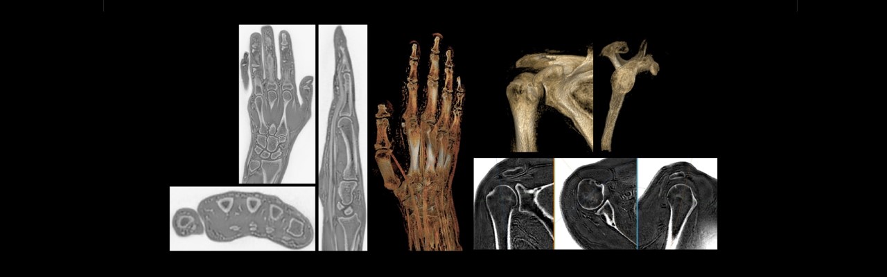
Specialty clinicians such as orthopedists were quick to realize the potential clinical applications for bone imaging with MRI for musculoskeletal (MSK) conditions. It is currently being utilized to obtain morphological information about the cortical bone structure in anatomies such as the shoulder, hip, skull, foot/ankle and spine. Its potential is especially welcome when longitudinal CTs are required, and particularly when low-dose CTs are unavailable. Having both soft tissue assessment and bone imaging within the same exam can also reduce the burden on the patient to attend two separate imaging modality appointments and may also lead to reduced healthcare costs.
Expanding the utility of MR capabilities
There are two types of bone tissue, trabecular bone and cortical bone. Traditional MRI is able to assess the trabecular, or internal compartment of bone tissue that consists of 25 percent bone and 75 percent marrow. Cortical or compact bone refers to the dense outer shell of bone tissue, which is approximately 90 percent bone and 10 percent pore space by volume. [1]
In MRI, bone is typically visualized with a void signal when using conventional clinical pulse sequences with echo times (TEs) of a several milliseconds or longer. The lack of direct signal originating from cortical bone impairs the ability of conventional MRI sequences to provide utility for assessing bone,[2] but using ZTE technology, it is now possible to image cortical bone surfaces. ZTE enables the acquisition of signal from tissues exhibiting the shortest T2 values.[3] ZTE imaging also has greater signal-to-noise ratio and scan-time efficiencies that are conducive to acquiring images at near-isotropic resolution and multiplanar reconstruction. The post-processing of ZTE images provides contrast between soft tissue and bone comparable to that at CT. [4]
Adding a 3 to 4 -minute ZTE sequence to an MRI acquisition does not interrupt the clinical workflow and provides clinicians with a one-stop shop for assessing soft tissue contrast in musculoskeletal (MSK) imaging and providing complementary cortical bone information that can be co-registered with the soft tissue series. The MRI bone imaging acquisition can be used across the patient’s entire anatomy.
“Many patients with injuries to the joints (e.g., shoulder and knee) or spine undergo multiple cross-sectional examinations (MR and CT) to assess soft and osseous tissues,” says Ryan Breighner, PhD, Computational Scientist at the Hospital for Special Surgery in New York. “For these patients, it may be possible to eliminate the need for a CT exam by adding one or more five-minute ZTE MR sequences to the end of their MR exam.”[5]
MRI-based qualitative imaging of cortical bone has drawn great interest among MSK radiologists and orthopedic and pediatric specialists. Expanding MRI capabilities to allow cortical bone visualization offers clinicians the opportunity to assess both tissue types simultaneously, which can help when planning treatments or special surgeries.

Improving access for vulnerable patient populations
While providing clinicians a simultaneous assessment of bone and the surrounding soft tissues, cortical bone imaging with MRI also reduces patient exposure to ionizing radiation if a separate CT is not required.[6]
“CT is typically used for bone imaging,” explains Eric Chang, MD, Professor of Radiology at the University of California San Diego, VA San Diego Healthcare System, in San Diego, California, “however, radiation is a concern. For example, in a CT of the spine, the neck, the thyroid will also get irradiated. If you image the thoracic or lumbar spine, other sensitive organs are irradiated, such as the breasts. In shoulder imaging, if you image one shoulder, both shoulders are irradiated, as well as the thyroid and parts of the breasts.”
Pathologic evaluation often requires imaging beyond standard radiography. MRI is often performed due to the modality’s inherent high soft-tissue contrast, ability to evaluate bone marrow, and non-ionizing radiation technique. MRI has gained utilization as an alternative to imaging with ionizing radiation when applicable, and especially for vulnerable patient populations such as pediatric and pregnant women.
“In pediatrics, CT is classically acquired to further evaluate cortical structures when needed,” explains Jesse Sandberg, MD, Professor of Pediatric Radiology at Lucile Packard Children’s Hospital, Stanford University School of Medicine in Stanford, California. “The drawback is ionizing radiation, which is obviously not ideal for pediatric patients. The application of an MRI sequence with cortical bone contrast would avoid ionizing radiation and may avoid the need for multimodal cross-sectional imaging.”
“We use the ZTE technique for diagnosis, presurgical planning and follow-up in our pediatric patients,” adds Ustun Aydingoz, MD, Professor of Radiology at Hacettepe University School of Medicine in Ankara, Turkey. The three main applications Dr. Aydingoz uses the ZTE technique include pediatric chest imaging, MSK imaging and neurological imaging, which includes the cranium, brain and sutures, and facial bones.
Growing opportunities in orthopedic care and surgical planning with oZTEo bone imaging
Cortical bone imaging with MRI can be utilized in a wide variety of MSK applications. As a MSK radiologist, Dr. Aydingoz uses the ZTE imaging technique in his routine clinical practice for MSK conditions and injuries such as traumatic, degenerative, inflammatory, and oncologic.
“We’ve incorporated ZTE into most of our protocols,” Dr. Aydingoz explains. “We also rely on it for critical shoulder angle measurement, for osseous bankart lesions, and for glenoid stock estimation and glenoid track assessment. We find the ZTE image is very much like the CT image and often, there’s no need to take an additional CT.”
Additionally, preoperative preparation with the use of 3D printed models is a growing area in orthopedics. The use of 3D–printed anatomic models has improved surgical planning, especially for patients in whom the conventional techniques are insufficient for establishing a proper strategy. [7] The quantitative information provided by ZTE regarding cortical bony structures, combined with soft tissue visualization can impact the intervention strategy. The extra information provided by 3D–printed models can lead to increased precision and impact patient risks, procedure times, and recovery times.[8]
Many patients with injuries to the joints are referred for MRI and CT imaging to assess soft tissues as well as bone structures. As more clinicians utilize ZTE for cortical bone imaging with MRI in a growing number of clinical applications, they will benefit from having both soft tissue assessment and bone imaging within the same exam to aid in diagnosis, surgical planning and follow-up, as well as keep their patients from additional exposure to ionizing radiation from other imaging studies.
RELATED CONTENT
- Read more about the clinical benefits of MR bone imaging, here.
- Explore more about GE Healthcare’s one-stop solution for orthopedic imaging with oZTEo, here.
DISCLAIMER
Not all products or features are available in all geographies. Check with your local GE Healthcare representative for availability in your country.
REFERENCES
[1] https://www.ncbi.nlm.nih.gov/pmc/articles/PMC5690546/
[2] https://www.frontiersin.org/articles/10.3389/fendo.2020.555756/full
[3] https://pubs.rsna.org/doi/full/10.1148/radiol.2017170906
[4] https://pubs.rsna.org/doi/full/10.1148/radiol.2017170906
[5] https://www.gesignapulse.com/signapulse/spring_2018/MobilePagedArticle.action?articleId=1396201#articleId1396201
[6] https://www.frontiersin.org/articles/10.3389/fendo.2020.555756/full#B26
[7] https://www.ncbi.nlm.nih.gov/pmc/articles/PMC6132335/
[8] https://www.ncbi.nlm.nih.gov/pmc/articles/PMC6132335/