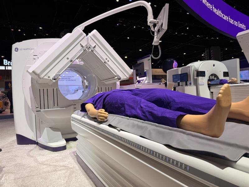
As the 2024 Radiological Society of North America (RSNA) Conference approaches, GE HealthCare remains steadfast in its commitment to delivering innovative imaging technology designed to address the unique needs of nuclear medicine departments. Whether it's improving image quality or enhancing workflow efficiency, every piece of technology – like the highly anticipated Aurora SPECT/CT system[i] – reflects insights from a range of stakeholders. Understanding the perspectives of department heads, radiologists, technologists, and physicists allows GE HealthCare to create solutions that meet the most pressing challenges faced by these professionals daily.
Each role within a nuclear medicine department evaluates technology differently based on their specific responsibilities. Department heads focus on clinical demands, operational economics, and scalability. Radiologists prioritize diagnostic confidence and imaging quality. Technologists value intuitive workflows and patient comfort, while physicists emphasize precision, and adaptability. GE HealthCare listens to all these voices to create scalable, impactful solutions that empower healthcare teams to deliver exceptional care.
Delivering advanced, patient-centered care for department heads
Patrick Flamen,[ii] head of Belgium’s comprehensive cancer center, must juggle clinical demands, staff productivity, and economic constraints, ensuring his department meets growing clinical demands while prioritizing patient safety and operational efficiency. Managing thousands of PET and SPECT/CT scans annually across three hospitals requires technology that helps enhance diagnostic precision while streamlining workflows and maintaining high patient volumes.
GE HealthCare’s approach to innovation incorporates solutions to help improve quality, enable reduction radiation dose, and optimize workflows. Solutions like the Aurora system, for instance, exemplify this. "Aurora has allowed us to provide more efficient care to our patients, especially those undergoing frequent scans,” this has been transformative.
The system’s dose-reduction capabilities have been invaluable for Flamen’s cancer patients, who often undergo multiple imaging procedures. Aurora’s ASIR-V enables reduction in CT dose by up to 80% compared to FBP at the same image quality.[iii] Reduction in CT dose is a critical for vulnerable individuals – including pediatric patients. Additionally, having a diagnostic CT helps save time by providing the necessary technology to skip trekking the patient to the CT department for other imaging needs. Aurora’s streamlined workflow and ease of use have allowed Flamen’s team to integrate the system quickly, cutting down on patient wait times.
Physicians value exceptional image quality and diagnostic efficiency
For radiologists like Carlos Artigas[ii] at the University Hospital of Brussels, technology must support precise diagnostics and help manage increasing workloads. Imaging solutions that offer impressive image quality and help speed up acquisition processes are essential. With Aurora, Artigas has found a versatile solution capable of handling various nuclear medicine applications, from bone scans to advanced ventilation-perfusion studies.
GE HealthCare’s innovations are designed with the aim of empowering physicians to review and assess images quickly and confidently. “The system’s versatility and real-time solutions have improved our diagnostic confidence while helping us manage patient volumes more effectively,” Artigas notes.
These technologies also provide radiologists with tools to simplify complex measurements, like kidney contouring during renograms, enabling timely clinical decisions. By addressing these needs, GE HealthCare helps physicians balance accuracy with efficiency.
Technologists rely on workflow efficiency and patient comfort
For Florence Mbougue,[ii] a medical imaging technologist at Erasme Hospital located in Bruxelles, Belgium, the right technology enhances both workflow efficiency and patient care. Technologists interact directly with patients, and intuitive systems like Aurora help simplify their work while ensuring a positive patient experience.
Physicists seek precision, safety, and adaptability
As a radiation physicist, Bruno Vanderlinden[ii] values imaging systems that deliver operational efficiency without compromising diagnostic quality. At Brussels University Hospital, Aurora has proven to be a multi-functional tool, seamlessly transitioning between planar, tomographic, and whole-body imaging.
Aurora is designed to provide impressive image quality, in addition to real-time monitoring and rapid transition capabilities has streamlined workflows across departments. For example, Vanderlinden’s team can quickly switch from one imaging mode to another, from a standard scan to a hepatobiliary scintigraphy, while ensuring each exam is conducted efficiently and precisely.
Shaping the future of nuclear medicine
Imaging technology must address the nuanced requirements of each professional in a nuclear medicine department. Whether it's delivering patient-centered solutions for department heads, diagnostic efficiency for physicians, intuitive workflows for technologists, or adaptability for physicists, GE HealthCare designs innovations that meet these diverse needs.
The Aurora SPECT/CT system exemplifies this commitment, offering efficient solutions that help advance diagnostic capabilities and confidence, which can make a meaningful difference across varied medical imaging settings and help redefine precision and efficiency.
To learn more about Aurora and GE HealthCare’s innovations, visit booth 7330 at the 2024 RSNA Conference in Chicago, IL, from December 1st – 4th, or visit the company’s virtual event center.
[i] Aurora is CE marked. Available for sale in EU countries. 510(k) pending at the US FDA. Not available for sale in the U.S and other non-EU countries.
[ii] The statements and experiences by individuals described here are based on his/her own opinions and on results that were achieved in his/her unique setting. Since there is no “typical” hospital/clinical setting and many variables exist, i.e. hospital size, case mix, staff expertise, etc. there can be no guarantee that others will achieve the same results.
[iii] a. ASiR-V reduces dose by 50% to 82% relative to FBP at the same image quality (Image quality as defined by low contrast detectability.)
b. In clinical practice, the use of ASiR‐V may reduce CT patient dose depending on the clinical task, patient size, anatomical location, and clinical practice. A consultation with a radiologist and a physicist should be made to determine the appropriate dose to obtain diagnostic image quality for the particular clinical task. Low Contrast Detectability (LCD), Image Noise, Spatial Resolution and Artifact were assessed using reference factory protocols comparing ASiR‐V and FBP. The LCD was measured using 0.625 mm slices and tested for both head and body modes using the MITA CT IQ Phantom (CCT183, The Phantom Laboratory), using a model observer method.