Cholecystectomy, the removal of the gallbladder, is one of the most common medical procedures in general surgery with over one million procedures worldwide each year.[i] The gallbladder stores bile to assist the body in breaking down food. Deposits can form in this area, also known as gallstones, and may cause gallbladder issues such as choledocholithiasis. In some cases, doctors may recommend the removal of the organ to treat these complications in hopes of relieving patients of the pain and discomfort caused by the gallstones.
Imaging modalities more commonly used for this procedure include intraoperative cholangiography (IOC) and indocyanine green (ICG). While these modalities can be effective and provide additional visualization, there are limitations to the visualization these methods provide. IOC and ICG are also considered time consuming, and can use invasive techniques.
As an example, IOC requires dye injections and utilizes an x-ray for images of the area, exposing individuals to radiation. Since patients are exposed to x-ray, the method can only be performed once and requires a radiographer to be present, resulting in further cost and time implications.
Elevating the standard of care
GE HealthCare is challenging the status quo, introducing a valuable tool for healthcare professionals. Intraoperative ultrasound (iUS) provides clear visualization of the anatomy with real-time diagnostic information to support surgeons with critical clinical decisions throughout the procedure. iUS is comparable to IOC for imaging cholecystectomies. Its non-invasive and non-radiating attributes allow surgeons to effectively visualize biliary and vascular anatomy. iUS has also proven to be a cost-efficient option designed for safer and efficient procedures.
With iUS:
- Surgeons have an option that provides real-time guidance and visualization of the anatomy, which can be used throughout the procedure as many times as needed.
- Surgeons can effectively detect common bile duct stones.
- Color flow doppler helps differentiate between vessels and ducts and is overall effective at imaging difficult cases.
- Patients and staff avoid exposure to radiation.
- There is no required pre-operative preparation.
|
|
|
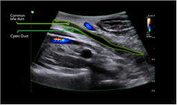
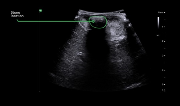
Images from laparoscopic cholecystectomy procedure using intraoperative ultrasound annotated to show stones and anatomy of interest during the procedure
“Without ultrasound, it would not have been possible to confirm the presence of stones in the bile duct, and I would not have been able to measure the diameter of the cystic duct and common bile duct,” said Mr. Somaiah Aroori, MB BS, MS (Surg), FRCS (Gen Surg), a consultant hepatobiliary and renal transplant surgeon at Derriford Hospital and University Hospital Plymouth. He emphasized how the continuous use of iUS provided useful information throughout the procedure and the real-time imaging made it possible to measure the size and number of stones to determine the best approach for removing the stones.
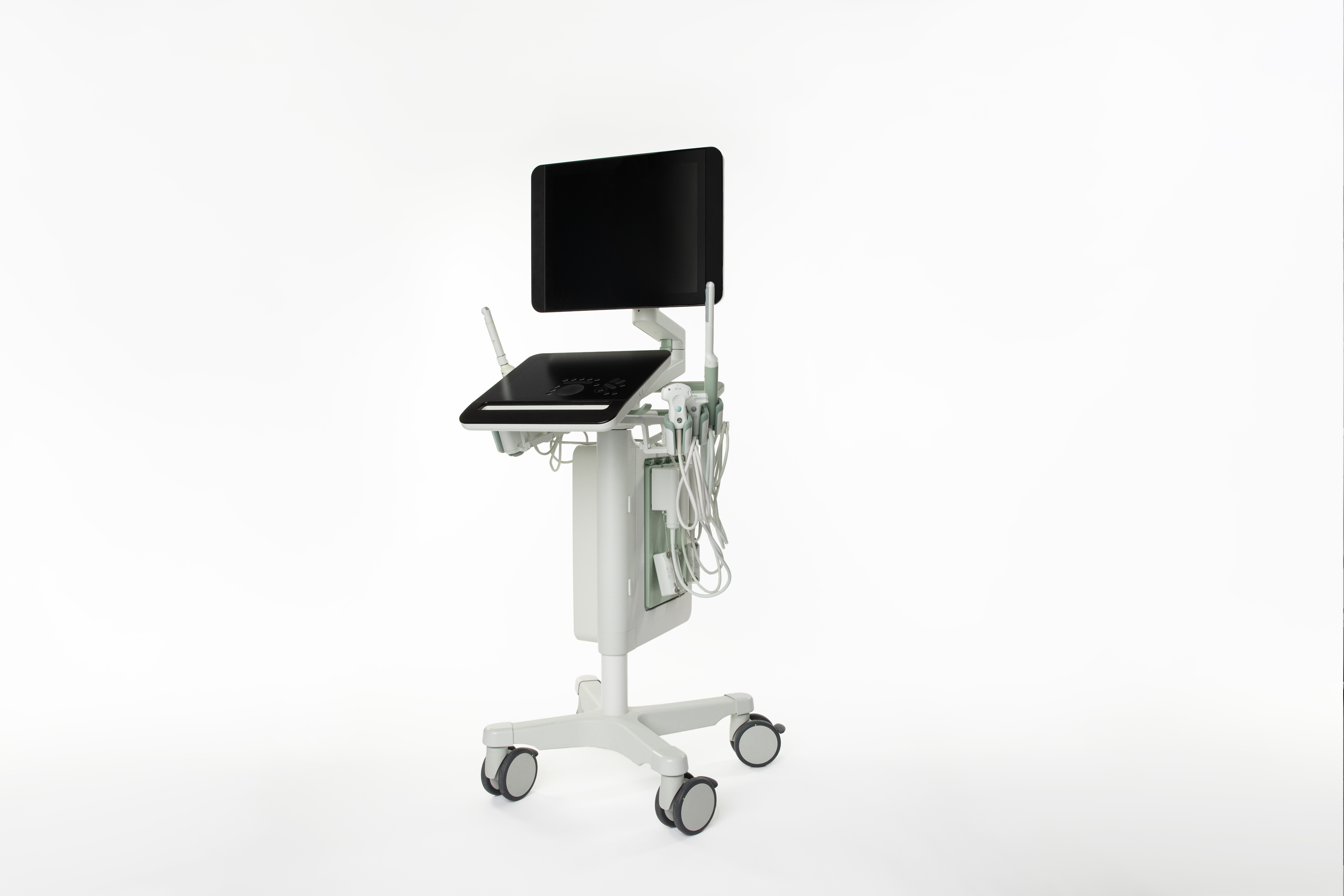
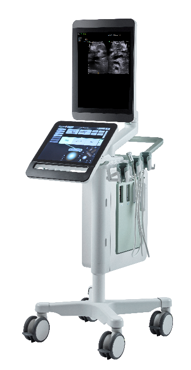
iUS options including bkActiv provide clinicians with critical information to make informed clinical decisions.
Learn about active imaging ultrasound solutions designed to reduce the iUS learning curve and elevate surgical procedures: https://www.bkmedical.com/applications/general-surgery-and-hepatobiliary-ultrasound-cholecystectomy/.
[i] Ambe P, Esfahani BJ, Tasci I, Christ H, Kohler L. Is laparoscopic cholecystectomy more challenging in male patients? Surg Endosc. 2011;25(7):2236–40.