By Sarah Handzel, BSN, RN
In terms of diagnostic capabilities and value, the ECG remains one of the best methods for identifying and accurately diagnosing a variety of cardiovascular conduction issues. Thanks to its low cost and relative ubiquity in healthcare settings, physicians often turn to ECG as an initial diagnostic tool. However, ECG lead misplacement continues to pose a challenge to workers in the healthcare industry, leading to potentially serious consequences like misdiagnosis and inappropriate or unnecessary treatment.
According to a recent study in Cardiology and Cardiovascular Medicine, some estimates show that nurses misplace ECG leads in up to 64% of all cases. The same study indicates that less than 20% of cardiologists correctly place V1 and V2 precordial electrodes.1 Misplacement of V1 or V2 may lead to false-positive findings, including acute anterior ST-segment elevation myocardial infarction or incomplete right bundle branch block.2
Clinicians of all experience levels should refresh their knowledge regarding ECG electrode placement to ensure accurate data capture that is reproduceable if another ECG is obtained. Adherence to professional standards and best practices for ECG lead placement both help clinicians retrieve the most accurate information possible without unnecessary delays or additions to treatment.
The Basics of Electrodes and Their Configuration
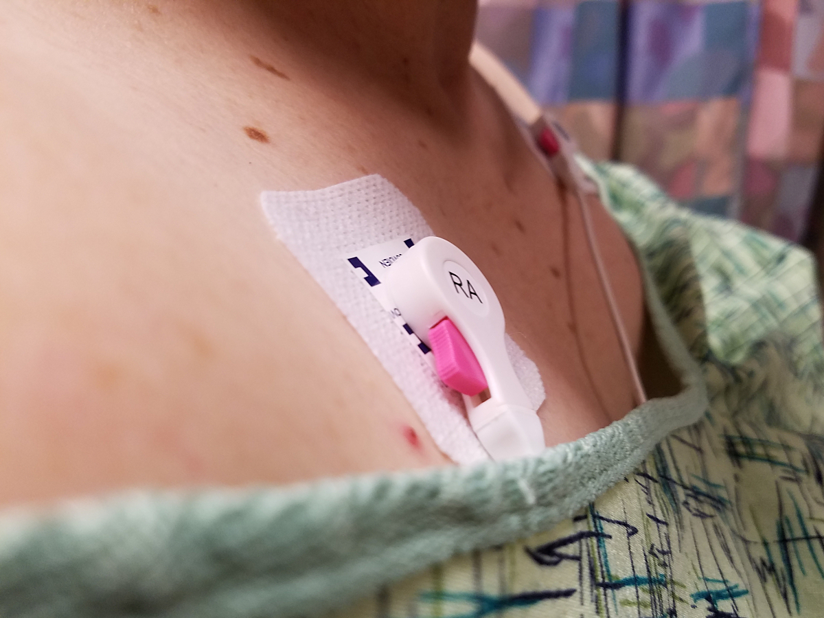
Any discussion of ECG lead misplacement warrants a brief review of electrodes. Most ECG electrodes are disposable; they may expire or dry out, and clinicians should check them before using to ensure neither has occurred. Ideally, all electrodes in a package should be used within the recommended time frame, as those left over in opened packages may lose functionality over time. Additionally, the electrodes used must match the type of ECG evaluation performed. For example, electrodes used with Holter monitors are different from those used for resting ECG. Clinicians should also only use one type of electrode for the entire ECG set-up instead of using multiple types of electrodes during a single exam.3
Regardless of the patient or suspected diagnosis, certain standards for ECG electrode placement should be followed. First, many people have oily skin—before applying any electrode, be sure to clean the placement area with an alcohol pad or soap and water and let it dry completely. If the patient is very hairy, it may be necessary to shave the site before applying the electrode.3
Limb electrodes should be placed first, followed by V1 and V2. The remaining electrodes must be placed correctly in the appropriate intercostal spaces. Clinicians should be sure to separate limb lead wires from chest lead wires. The limb lead wires are usually longer and are located on the outside of each lead.3
For procedures, such as prolonged 12 lead monitoring or 12 lead Holter, while the chest electrode configuration remains the same, electrodes placed on the limbs are moved onto the patient's torso. Both the right and left arm electrodes should be placed in the intraclavicular fossae, approximately two centimeters below the clavicle. The right and left leg electrodes remain on the anterior axillary line, halfway between the iliac crest and the costal margin. This Mason Likar electrode configuration is also used for stress testing, and clinicians should remember that such placement may show variations from normal ECGs. It is not uncommon for ECG waveforms to demonstrate morphological variations such as obscured Q waves in inferior leads.4

Examples of Misplacement and Impact on Diagnosis
Proper placement of electrodes ensures that any ECG recorded will be as accurate as possible. When electrodes are misplaced, the potential impacts on the entire diagnostic and treatment process may be significant.
Misplacement of limb leads is a common problem that produces various pseudo results. Unintentional reversal of limb leads may produce results suggestive of lateral infarct, inferior infarct, or negative QRS/T complexes. Because each limb lead records the cardiac axis in the frontal plane, axis abnormalities may also indicate misplacement. These abnormalities may include extreme QRS axis, abnormal P wave axis, inverted lateral limb leads, concordantly negative complexes in all limb leads, or low or isoelectric voltages isolated in a single limb lead.5
For example, misplacement of the right and left arm electrodes may produce an ECG featuring both Q wave and T wave inversion in leads I/aVL. The P wave would be negative in lead I but positive in lead aVR. Clinicians may suspect a lateral infarct when no such problem exists. Or, reversing the left arm and left leg electrodes results in an ECG with negative QRS/T complex in lead III together with a negative P wave.5
Precordial lead misplacement is also a known issue among clinicians. Depending on the lead affected, cardiologists may see early R wave progression or a pattern suggesting a pseudo-infarction. And even if all precordial leads are placed in the correct sequence, placing leads V1 and V2 too high on the patient's chest can result in various incorrect ECG patterns suggestive of complications like right bundle branch block, ST-segment elevation, or anterior T wave inversions. Cardiologists may interpret these false results as evidence of cardiac ischemia or pulmonary embolism.5
Patient-specific Considerations When Placing ECG Electrodes
Certain patient factors may influence the accuracy of information captured during a standard 12-lead ECG. Adults commonly lose body mass as they age—electrodes should never be placed over bony prominences on any older patient. Bones many interfere with wave transmission.
Similarly, artifact may appear if a clinician places electrodes over large muscles or skin folds. In the case of obese patients, providers should be sure to move large skin folds to expose common electrode application sites. This is also true for women with extensive breast tissue.6
Patients in respiratory distress may cause significant artifact on standard ECGs. Artifact is typically more common in the chest or precordial leads, but muscle artifact is also possible if the chest sinks in below the chest with each inhalation.7
Although ECG lead misplacement is relatively common, implementing best practices for electrode placement helps mitigate possible errors. A review of best practices is a good way for clinicians to brush up their skills so they may obtain the most accurate ECG readings possible.

Resources:
1. Hadjiantoni A, Oak K, Mengi S, Konya J, Ungvari T. Is the correct anatomical placement of the electrocardiogram (ECG) electrodes essential to diagnosis in the clinical setting: a systematic review. Cardiology and Cardiovascular Medicine. 2021;5(2):182-200. https://www.fortunejournals.com/articles/is-the-correct-anatomical-placement-of-the-electrocardiogram-ecg-electrodes-essential-to-diagnosis-in-the-clinical-setting-a-syste.html
2. Abobaker A, Rana RM. V1 and V2 pericordial leads misplacement and its negative impact on ECG interpretation and clinical care. Annals of Noninvasive Electrocardiology. 2021;26(4). https://www.ncbi.nlm.nih.gov/pmc/articles/PMC8293594/
3. Diagnostic ECG Lead Placement. GE HealthCare. https://clinicalview.gehealthcare.com/poster/diagnostic-ecg-lead-placement. Accessed April 21, 2023.
4. The role of optimal and modified lead systems in electrocardiogram. Advanced Methods in Biomedical Signal Processing and Analysis. https://www.sciencedirect.com/science/article/abs/pii/B9780323859554000144. Accessed June 20, 2023.
5. ECG Cases 29 Misdiagnosis from Lead Misplacement, Artifact and Lead Reversal. Emergency Medicine Cases. https://emergencymedicinecases.com/ecg-cases-lead-misplacement-artifact-lead-reversal/. Accessed May 10, 2023.
6. Best Practices for ECG Lead Placement on Women. GE HealthCare. https://www.gehealthcare.com/insights/article/best-practices-for-ecg-lead-placement-on-women. Accessed April 21, 2023.
7. "Performing High Quality Ecgs on Patients in Respiratory Distress." Performing High Quality ECGs on Patients in Respiratory Distress. GE HealthCare, 2020. https://www.gehealthcare.com.tr/-/jssmedia/global/products/files/diagnostic-ecg/dcar-global-lead-placement-respiratory-distress-final-jb77439xx.pdf?rev=-1.