Magnetic Resonance Imaging (MRI) technology is widely used in healthcare today and has transformed clinical diagnostics with non-invasive and non-ionizing radiation imaging techniques. Its many benefits make it a critical tool in clinical applications such as cardiology, where MRI delivers high-resolution visualization of myocardial structure and function and provides characterization of myocardial tissue. The introduction of artificial intelligence (AI) and deep learning algorithms for imaging reconstruction is an exciting realization of advanced technology that is enabling faster, more intelligent diagnostic imaging systems such as MR to support clinical decision-making.
AI is helping to advance medical imaging
The use of AI and deep learning technologies are firmly established as essential tools for progress in many healthcare applications. Specifically, within medical imaging, over 500 AI-enabled devices have been approved for clinical use by the US Food and Drug Administration (FDA).[1] Deep learning, a subset of machine learning, has become an integral part of AI solutions that are developed for medical imaging applications. Applying deep learning algorithms to MR image reconstruction enables improvements in MR that haven’t been possible using traditional reconstruction methods. Most recently, deep learning-enabled MR image acceleration solutions for cardiovascular imaging applications are revolutionizing the capability to image the beating heart.
“Our vision is to image at the speed of life,” said Dr. Anja Brau, General Manager of MR Clinical Solutions and Research Collaborations at GE HealthCare. “GE HealthCare is very proud to lead the industry in AI-enabled medical device authorizations, and we continue to fulfill our commitment to infusing AI into our products with this next chapter in our deep learning revolution for MR. There’s no application that is more demanding than cardiac imaging, and we challenged ourselves to develop a deep learning solution to image at the speed of the human heart.”
According to the National Institute of Health (NIH), coronary artery disease is still the leading cause of death in the US, and the third leading cause of death worldwide, even with improvements in therapeutic and interventional methods.[2],[3] Therefore, the importance of clinicians’ ability to visualize the structure and function of the heart and heart tissue cannot be overstated. MR is considered the gold standard for diagnostic cardiac imaging[4], but acquisition can require lengthy amounts of time, and frequent breath holding, which is stressful on some patients, where others are unable to do it at all.
Cardiac imaging is also complicated by motion—motion from the movement of the patient during free breathing and motion from the heartbeat itself. Applying deep learning algorithms to MR image acceleration can help to overcome the historical trade-offs between scan time and image quality, especially when patients have difficulty holding their breath or lying still for long periods of time.
Enhancing MR scan speed with deep learning reconstruction
GE HealthCare initially introduced AIR™ Recon DL, a deep learning trained algorithm to achieve higher signal-to-noise ratio (SNR) by leveraging all of the raw MRI acquisition data to reconstruct sharper and accurate images. This application has enabled compelling results and has been used in more than 19 million patient exams thus far.*
“Even so, conventional cardiac MRI is just still too slow to capture all the frames that you need across a heartbeat,” explained Dr. Brau, “so for decades, clinicians imaged portions of data across multiple heartbeats, and those data were stitched together. However, it requires the patient to hold their breath to avoid motion artifacts. And that’s just for one slice. Twelve breath holds are needed for the whole short axis, but you also need to image the long axis, and so on. This is incredibly demanding for the patient and for the technologists.”
To solve the motion limitations of MR cardiac imaging, GE HealthCare developed Sonic DL™, its next chapter in deep learning reconstruction for MR, designed to provide highly accelerated functional imaging of the heart. This deep-learning reconstruction algorithm allows for the unprecedented ability to perform MR scans within a single heartbeat, while having sufficient resolution to enable measurement of blood volume and substantially reduce the amount of time it takes to obtain the images needed for diagnostic evaluation.
This deep-learning reconstruction solution is capable of up to twelve times acceleration** and allows the conventional acquisition, which took about ten heartbeats (R to R), to be completed in just one heartbeat, and in three heartbeats for slightly higher resolution.
Impacting clinical care with cardiac imaging speed
Used in clinical practice, it’s having a tremendous impact on cardiovascular imaging for patients.
“We started our journey with deep learning image reconstruction prior to having Sonic DL™ available,” said Dr. Melany Atkins, Division Chief of Body/Cardiovascular Imaging and Medical Director of Advanced Cardiac Imaging at Inova Health System and Fairfax MRI Center in Fairfax, Virginia. “This new deep learning application afforded us the ability to do rapid single beat acquisition coverage of the entire ventricle in one to two breath holds for our non-C sequences. We also added a gray blood sequence, which allows us to identify those subtle sub-endocardial delayed enhancements.”
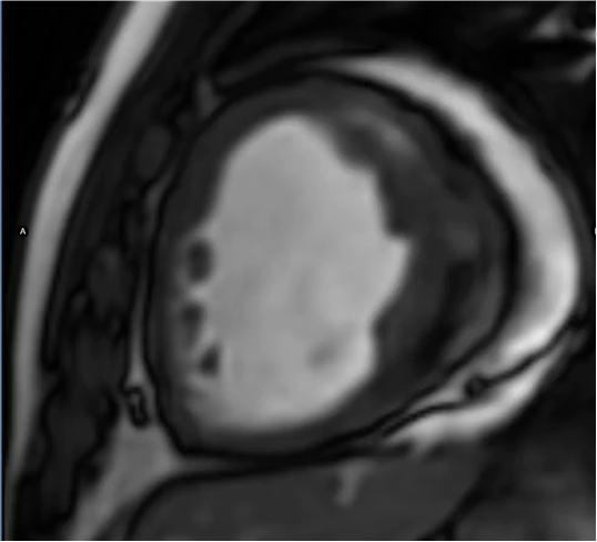
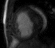
In a comparison of images from a 14-year-old patient with congenital heart disease, Dr. Atkins summarized the differences between recent imaging using Sonic DL™ and prior imaging without it.
 |
 |
|
Sonic DL™ 3 R to R |
Conv SSFP Same patient, 2 years prior |
“This patient is unable to hold his breath and is also claustrophobic, so it’s difficult for him to comply with the exam. It’s easy to appreciate the very poor image quality on the prior exam compared to our current study using Sonic DL™, and certainly a significant time savings.”
According to Dr. Atkins, many of her patients can comply with minimal breath hold requirements, but she is looking forward to utilizing the new imaging technology to improve cardiac MR for many more patients.
“We’re fortunate that in our mainstream outpatient imaging center, our patients are often able to hold their breath a little bit. So that’s why we’ve continued to do Sonic DL™ at three heartbeats, just to have the improvements in SNR and faster temporal resolution. It’s probably 25 percent or less of our patients who really need a full free-breathing exam, but that’s probably going to increase as time goes on. Now that it’s available to do, everybody will want to have the free-breathing exam,” Dr. Atkins said.
The future of MRI with AI innovation
Advances in MRI technology, as well as AI-based innovations in image reconstruction, such as GE HealthCare’s AIR™ Recon DL and Sonic DL™ have enabled compelling results. As these solutions become more widely utilized across different clinical applications such as cardiology, they can provide clinicians with new insights into diseases and treatments, as well as improve the patient experience.
RELATED CONTENT
- View the exclusive presentation, now on-demand from RSNA 2023: Setting the pace for innovation in MR with deep learning
- View on-demand: Breaking barriers in MR with deep learning
DISCLAIMERS
Not all products or features are available in all geographies. Check with your local GE HealthCare representative for availability in your country.
*Calculated by IB data with estimation 20 scans per day, 5.5 working day in a week, fully start using AIR™ Recon DL 4 weeks after delivery, as of January, 2024.
** Data on file, 2023, Sonic DL™ vs fully sampled CINE acquisition.
[1] https://healthexec.com/topics/artificial-intelligence/fda-has-now-cleared-more-500-healthcare-ai-algorithms
[2] Saeed M, Van TA, Krug R, Hetts SW, Wilson MW. Cardiac MR imaging: current status and future direction. Cardiovasc Diagn Ther. 2015 Aug;5(4):290-310. doi: 10.3978/j.issn.2223-3652.2015.06.07. PMID: 26331113; PMCID: PMC4536478.
[3] Brown JC, Gerhardt TE, Kwon E. Risk Factors for Coronary Artery Disease. [Updated 2023 Jan 23]. In: StatPearls [Internet]. Treasure Island (FL): StatPearls Publishing; 2023 Jan-. Available from: https://www.ncbi.nlm.nih.gov/books/NBK554410/
[4] Piersson, A.D. Essentials of cardiac MRI in clinical practice. J Cardiovasc Magn Reson 18 (Suppl 1), T10 (2016). https://doi.org/10.1186/1532-429X-18-S1-T10

