About 60 percent of patients with cancer need radiation therapy,[1] which requires high-quality medical imaging. The use of medical imaging to guide radiation therapy facilitates improved treatment efficiencies by more accurately tracking tumor response so that treatment may be adjusted accordingly. As imaging technologies have become more advanced, they offer clinicians information not only about the size of the lesion before, during and after treatments, but also about the tumor’s tissue characterization and metabolic activity.
While various imaging modalities such as magnetic resonance imaging (MRI) and positron emission tomography (PET) are often used in conjunction with computed tomography (CT) to provide these types of information for clinicians, current practice uses CT as the standard imaging modality for radiotherapy planning, because it provides a three-dimensional view of the tumor as well as data regarding tumor density, which is used for dose calculations in radiotherapy planning.[2] Traditional CT imaging data drives the current practice of delivering a relatively homogeneous radiation dose to the entire clinical target volume of the tumor.[3]
The development of artificial intelligence (AI)-based tools, however, is making effective radiation treatment planning possible without a heavy reliance on CT, supporting the emerging use of other imaging modalities, such as magnetic resonance imaging (MRI) for effective treatment planning and monitoring, as well as helping to improve tumor dose calculations, based on tumor biology.
Advanced medical imaging enables adaptive radiotherapy planning
The use of medical imaging throughout the course of a patient’s radiation treatment enables any anatomical changes in lesions to be detected so that treatment plans can be changed as necessary. This type of adaptive radiation therapy planning--where an individual plan can be created for each treatment fraction, depending on the precise positioning, and shape of the tumor at the time of imaging--is an opportunity for clinicians to help improve patient outcomes through the use of functional imaging techniques that can accurately map tumor characteristics such as hypoxia, vascularity, and cellular proliferation.
Supporting this process, the application of functional imaging techniques has shed light on the importance of understanding tumor biology and could help identify areas of radio-resistance within a tumor that require radiation dose escalation.[4] By enabling a greater understanding of tumor biology, functional imaging techniques such as PET/CT, or perfusion and kinetic perfusion studies can provide visibility to tumor characteristics at a metabolic level. Traditional CT imaging without the additional functional information can be challenging when delineating tumors from surrounding soft tissue. In certain applications, other imaging modalities are preferred. For example, MRI provides higher resolution and greater soft tissue contrast in pelvic tumors when compared to CT imaging, and PET imaging with fluorodeoxyglucose (FDG) provides more accurate tumor delineation in head and neck, lung, lymphoma and esophageal cancers.[5],[6],[7],[8]
Advances in MRI techniques include quantitative applications that have expanded its use in radiation oncology, not only because they can now provide functional information on tumor tissues, but also because MRI does not expose patients to ionizing radiation.
The emergence of AI within radiation oncology treatment planning
Because of radiation oncology’s heavy reliance on digital data processing for accurate radiation therapy planning, AI is a natural fit. AI applications have the potential to provide a quantitative assessment of clinical conditions and improve the accuracy, precision, efficiency, and overall quality of radiation therapy for patients with cancer.[9]
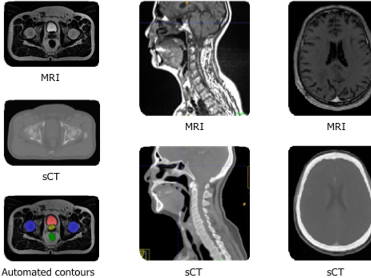
AI tools and deep learning (DL) applications are helping radiation oncologists to integrate and co-register patient data, as well as to automate many therapy planning tasks. New AI tools in MRI are helping radiation oncologists in therapy planning with a new ability to build synthetic CT images from MRI, supporting the emerging trend of MRI-only radiation therapy.
Using a DL image reconstruction algorithm and making full use of the raw data from MRI scans to reduce image noise, maximize image quality and resolution, high-quality MR images are converted to synthetic CT images. Clinicians can rely on both sets of images for accurate tumor volume and soft tissue delineation, eliminating the need for a conventional CT examination, as well as the work involved in multimodal image registration. This innovation streamlines the clinical workflow and allows for more precise treatment delivery.
The future of precision radiation oncology
The integration of advanced imaging technology into radiotherapy treatment planning and radiotherapy treatment systems has led to an increase in the precision and accuracy of radiation delivery.[10] The continued development of sophisticated AI tools and DL techniques in medical imaging will only add to the ability of clinicians to more accurately and precisely characterize and target tumors with appropriate therapies.
Learn more about MR radiation oncology solutions.
Learn more about Radiation Therapy Planning solutions.
Read the associated press release for more information: GE Healthcare to Offer End-to-end Deep Learning Solutions with Spectronic Medical to Improve Accuracy for Radiation Oncology Treatment.
REFERENCES
[1] NIH National Cancer Institute, Radiation Therapy and You: Support for People with Cancer, NIH Publication No. 17-7157, October 2016.
[2] Bhide, S. A., Newbold, K. L., Harrington, K. J. & Nutting, C. M. Clinical evaluation of intensity-modulated radiotherapy for head and neck cancers. Br. J. Radiol. 85, 487–494 (2012).
[3] Grégoire, V. & Haustermans, K. Functional image-guided intensity modulated radiation therapy: integration of the tumour microenvironment in treatment planning. Eur. J. Cancer 45, 459–460 (2009).
[4] Beaton, L., Bandula, S., Gaze, M.N. et al. How rapid advances in imaging are defining the future of precision radiation oncology. Br J Cancer 120, 779–790 (2019). https://doi.org/10.1038/s41416-019-0412-y.
[5] Daisne, J. F., Duprez, T., Weynand, B., Lonneux, M., Hamoir, M. & Reychler, H. et al. Tumor volume in pharyngolaryngeal squamous cell carcinoma: comparison at CT, MR imaging, and FDG PET and validation with surgical specimen. Radiology 233, 93–100 (2004).
[6] . MacManus, M., Nestle, U., Rosenzweig, K. E., Carrio, I., Messa, C. & Belohlavek, O. et al. Use of PET and PET/CT for Radiation Therapy Planning: IAEA expert report 2006–2007. Radiother. Oncol. 91, 85–94 (2009).
[7] Ashamalla, H., Rafla, S., Parikh, K., Mokhtar, B., Goswami, G. & Kambam, S. et al. The contribution of integrated PET/CT to the evolving definition of treatment volumes in radiation treatment planning in lung cancer. Int. J. Radiat. Oncol. Biol. Phys. 63, 1016–1023 (2005).
[8] Viswanathan, A. N., Dimopoulos, J., Kirisits, C., Berger, D. & Potter, R. Computed tomography versus magnetic resonance imaging-based contouring in cervical cancer brachytherapy: results of a prospective trial and preliminary guidelines for standardized contours. Int. J. Radiat. Oncol. Biol. Phys. 68, 491–498 (2007).
[9] Huynh, E., Hosny, A., Guthier, C. et al. Artificial intelligence in radiation oncology. Nat Rev Clin Oncol 17, 771–781 (2020). https://doi.org/10.1038/s41571-020-0417-8.
[10] Jaffray, D. A. Image-guided radiotherapy: from current concept to future perspectives. Nat. Rev. Clin. Oncol. 9, 688–699 (2012).