Advances in Positron Emission Tomography (PET) support its continued use as an important clinical tool. PET is actively used in a diverse range of applications including in oncology, cardiology, and neurology. It also utilizes radiotracers such as 18F-based PET tracers and FDG to enable visualization and measurement of a number of biological processes. Understanding how the human body metabolizes FDG and other radiotracers offers clinicians insights. Quantitative imaging of physiological, biochemical and pharmacological targets have paved the way for such applications as the metabolic assessment of tumors and quantification of tumor processes.
As PET technology continues to evolve, sophisticated digital detectors and image reconstruction software enable high levels of sensitivity, image quality, and diagnostic insights that inform clinical decisions. Traditional clinical use of PET technology employs a static approach to the image acquisition, where imaging is acquired at a single time frame, after the patient is injected with the radiotracer.[1] New imaging methods are currently utilized to image the patient using a dynamic, whole-body approach to PET imaging.
Strategic Business Leader for Molecular Imaging at GE Healthcare
Improving the visualization of disease processes with parametric PET imaging
Whole-body dynamic PET imaging presents not only the opportunity to visualize the uptake of radiotracer in patients, but also presents the opportunity to understand tracer distribution and the metabolic process that is occurring as the patient is being imaged. Understanding disease through the analysis of dynamic whole-body PET imaging, also called dynamic PET or parametric pet imaging, is the next frontier in molecular imaging. Research continues and clinical utilization has begun. Industry leaders are working collaboratively with clinical experts to understand the potential clinical applications for parametric PET imaging and how it might move into routine clinical utilization.
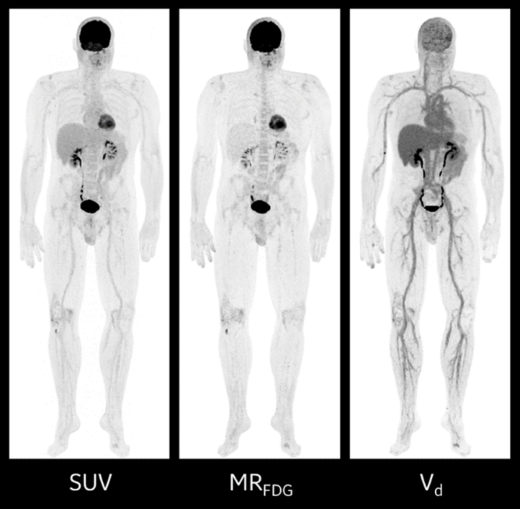
Standard PET imaging yields three-dimensional scans that capture the spatial resolution of a radiotracer at a single point in time. The images are usually quantified using a standardized uptake value (SUV) that captures that distribution.[2] Image analysis is often qualitative, with clinicians providing their impressions, supported by semi-quantitative analysis using the SUV.[3]
Dynamic PET imaging may require additional acquisitions but has the advantage that it measures the full spatiotemporal bio-distribution of a radiotracer. In combination with tracer kinetic analysis, it enables the generation of parametric images that more directly quantify underlying biological parameters of interest, such as blood flow, glucose metabolism, and receptor binding.[4]
While performance of parametric imaging has been limited by noise, the increased sensitivity of newer scanners, in combination with variable acquisition and analysis protocols, and advanced image reconstruction algorithms can further improve the data quality of dynamic PET for parametric imaging,[5] to facilitate a move into routine clinical utilization.
Using advanced PET imaging for PET parametric imaging
Dedicated whole-body PET scanners are not a prerequisite for dynamic imaging for whole-body parametric images of metabolic activity and multi-bed and multi-pass parametric imaging, as this can equally be performed with existing PET scanners. Hardware has experienced dramatic improvements in improving intrinsic scanner sensitivity, which can result in improved image quality for parametric images derived from kinetic modeling.[6] And the axial field of view (AFOV) has increased greatly, making it even easier to accommodate the dynamic whole-body protocols without the need for a dedicated whole-body scanner. The scan times required for dynamic whole-body PET imaging may increase the potential for patient movement, respiratory motion, and cardiac motion,[7] but powerful image reconstruction solutions are helping to reduce any artifacts they may cause.
Using advanced PET imaging and novel imaging biomarkers, whole-body dynamic PET imaging of radiotracer expression can be quantified and used for optimizing and predicting therapy response in oncology and for assessment of inflammation. Parametric images also have the potential for improved detection due to their high contrast and for more accurate and earlier therapeutic response assessment.
Martin Hüllner, PD, MD, Senior Physician, Department of Nuclear Medicine, and Deputy Clinic Director, Department of Nuclear Medicine at University Hospital in Zurich, Switzerland, shared some of his experiences with whole-body dynamic PET imaging. “Why and how are we doing whole-body dynamic PET imaging?” Dr. Hüllner said. “It’s a technique that’s going from clinical research to clinical routine.”
Dr. Hüllner explained that while static PET imaging using SUV is the most commonly used quantitative measure for tumor detection, staging and for therapy response assessment, there are factors that can have a major influence on clinical SUV accuracy and variability. “Erroneous SUV measurements,” he says, “may result in continuation of ineffective therapy, or in the discontinuation of effective therapy, so this is a major problem in clinical routine.”
Using GE Healthcare’s Discovery™ MI Gen 2 PET/CT scanner, Dr. Hüllner uses two different imaging protocols to complete the whole-body dynamic PET imaging exams in his facility.
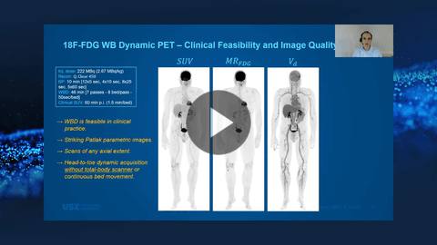
“The scanner has a sensitivity of approximately 30 counts per second per kilobecquerel and an axial field of view of 30 cm,” he explained. “During the acquisition, it’s actually very convenient in the system, you can select the acquisition time for each bed and for each pass. This can be done very conveniently on the console using the user interface,” he continued. “The parametric images have a striking quality. You can do dynamic scans of any axial extent, and very important, you can do a head-to-toe dynamic acquisition without the need for a total body scanner, and without the need for continuous bed movement. So, using only the 30 cm axial coverage, you can do whole-body dynamic imaging.”
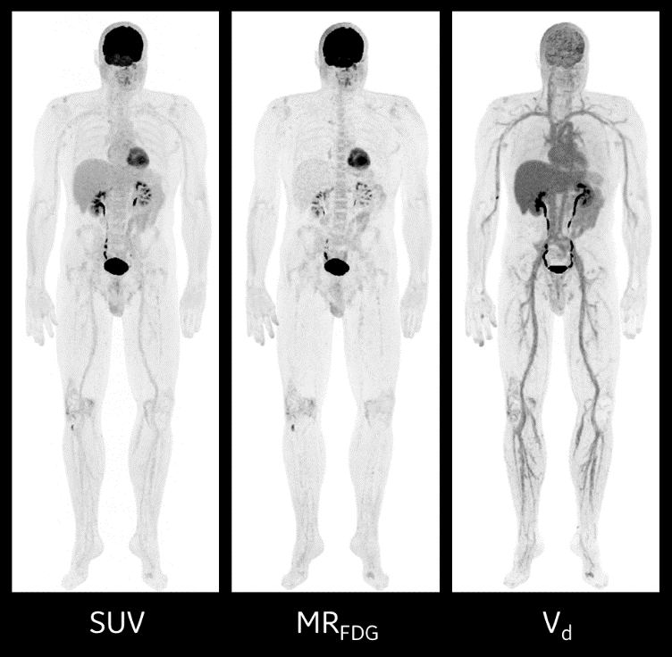
Example Whole-body Dynamic PET/CT*
FDG Inj. dose: 222 MBq (6 mCi), 2.67 MBq/kg
WBD Acquisition: 7 passes, 50 sec/bed; SUV: 1.5 min/bed, 60 min post inj.
Visualizing the future of molecular imaging with whole-body dynamic PET
Experts predict that dynamic PET imaging will be well-suited to study single organs and evaluate organ-specific diseases.[8] Neurological and cardiovascular studies have been the initial areas of focus in single bed dynamic studies till recently, but extending to whole-body dynamic acquisition, other areas of interest for potential clinical applications include the lungs, liver and other target regions in oncology applications.
Industry leaders such as GE Healthcare will continue to work with clinicians as they determine the most suitable clinical applications for whole-body dynamic PET imaging and will continue to bring forward innovative solutions to enable these new applications to be used in routine clinical care.
Department of Nuclear Medicine, University Hospital Zurich, University of Zurich, Switzerland
Whole-body dynamic PET/CT: From clinical research to clinical routine
Learn more about Discovery™ MI Gen 2, here.
With Discovery MI Gen 2 and Whole Body Dynamic IQ Protocol[9], you have a powerful new tool that provides you with the ability to better identify regions of metabolic activity and increased tracer uptake rate.[10]

DISCLAIMER
*Dynamic IQ application is not cleared or approved in the US. Not commercially available in the US.
Not all products or features are available in all geographies. Check with your local GE Healthcare representative for availability in your country.
REFERENCES
[1] Wang G, Rahmim A, Gunn RN. PET Parametric Imaging: Past, Present, and Future. IEEE Trans Radiat Plasma Med Sci. 2020;4(6):663-675. doi:10.1109/trpms.2020.3025086
[2] Wang G, Rahmim A, Gunn RN. PET Parametric Imaging: Past, Present, and Future. IEEE Trans Radiat Plasma Med Sci. 2020;4(6):663-675. doi:10.1109/trpms.2020.3025086
[3] Rahmim, A., Lodge, M.A., Karakatsanis, N.A. et al. Dynamic whole-body PET imaging: principles, potentials and applications. Eur J Nucl Med Mol Imaging 46, 501–518 (2019). https://doi.org/10.1007/s00259-018-4153-6
[4] Wang G, Rahmim A, Gunn RN. PET Parametric Imaging: Past, Present, and Future. IEEE Trans Radiat Plasma Med Sci. 2020;4(6):663-675. doi:10.1109/trpms.2020.3025086
[5] Wang G, Rahmim A, Gunn RN. PET Parametric Imaging: Past, Present, and Future. IEEE Trans Radiat Plasma Med Sci. 2020;4(6):663-675. doi:10.1109/trpms.2020.3025086
[6] Karakatsanis NA, Lodge MA, Tahari AK, Zhou Y, Wahl RL, and Rahmim A, “Dynamic whole body PET parametric imaging: I. Concept, acquisition protocol optimization and clinical application,” Phys. Med. Bio, vol. 58, no. 20, pp. 7391–7418 2013.
[7] Gallezot JD, Lu YH, Naganawa M, and Carson RE, “Parametric Imaging With PET and SPECT,” IEEE Transactions on Radiation and Plasma Medical Sciences, vol. 4, no. 1, pp. 1–23, 2020.
[8] Wang G, Rahmim A, Gunn RN. PET Parametric Imaging: Past, Present, and Future. IEEE Trans Radiat Plasma Med Sci. 2020;4(6):663-675. doi:10.1109/trpms.2020.3025086
[9] Whole-body Dynamic IQ Protocol enhances acquisitions currently limited to single FOV dynamic acquisition or manual prescription of multiple whole-body static scans.
[10] Processing software is needed for diagnostic purposes.