Advancements in technology, especially AI, have the potential to reshape the field of diagnostic imaging by improving accuracy, speed and image clarity. These tools are supporting clinicians in helping cancer patients receive the right treatments sooner and enabling clinicians to provide the best possible care.
“Radiologists read and interpret a number of diagnostic imaging studies: X-ray, CT, ultrasound, MR, and PET,” says Dr. Lloyd Stambaugh, president of Evergreen Radia Imaging Center, in Kirkland, Washington, U.S., and vice-president of Radia PS. “Our job is to read the studies and make a better diagnosis.”
AI and related technologies are empowering clinicians and imaging technicians in multiple ways. AI-enabled tools can automate repetitive tasks and transform unstructured medical data into meaningful insights, alleviating stress on healthcare professionals. Efficiency gains are especially crucial for radiology technologists; 18% of these positions were unfilled in the U.S. in 2023, lengthening patient wait times and intensifying the pressure on existing staff.
Two branches of precision cancer care — molecular imaging and cancer immunotherapy — are also benefiting from advances in imaging technology. AI-driven molecular imaging is delivering more detailed pictures of different types of cancer cells to help identify the therapies best suited for each patient and monitor progress throughout treatment. Advanced diagnostic imaging is having a similar effect on immunotherapy: With more than 5,000 drugs in development, understanding each patient’s immune response to specific drugs is key to improving individual outcomes — and providing big-picture insights that will benefit healthcare for decades to come.
Evergreen Radia Imaging Center uses Precision DL software, an AI-based method for processing positron emission tomography (PET) and computed tomography (CT) scans. “Precision DL takes the current state of PET/CT and makes it even better,” Dr. Stambaugh says.
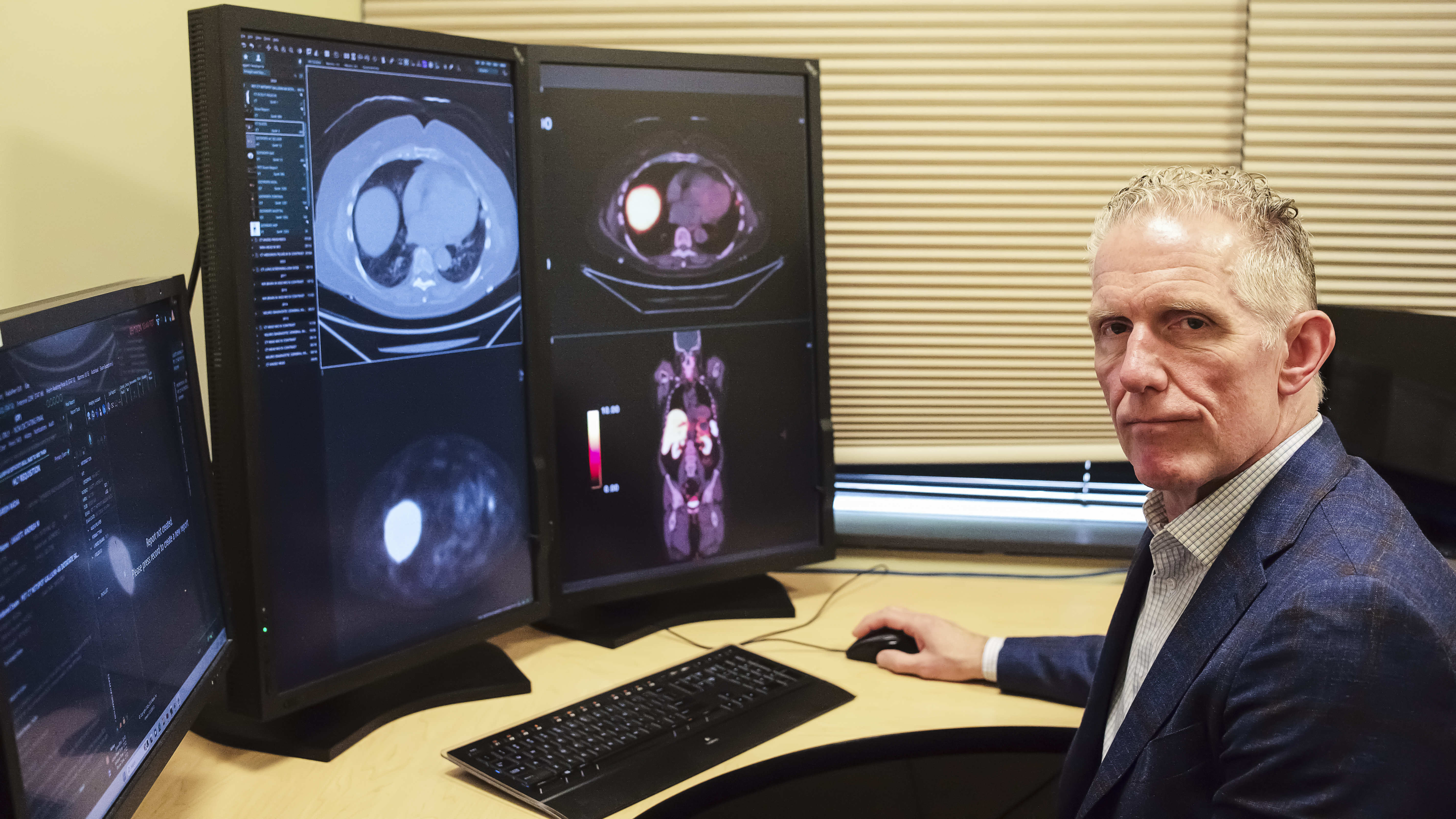
Dr. Lloyd Stambaugh
Until recently, for example, without AI assistance PET/CT scanners could not identify the spread of cancer to lymph nodes smaller than 8 millimeters in diameter*. “Finding early spread of cancer to lymph nodes or to other parts of the body… may make the difference in weeks or even months of the patient being able to receive treatment earlier,” Dr. Stambaugh says. “The earlier a cancer is diagnosed, the better chance the patient has of surviving.”
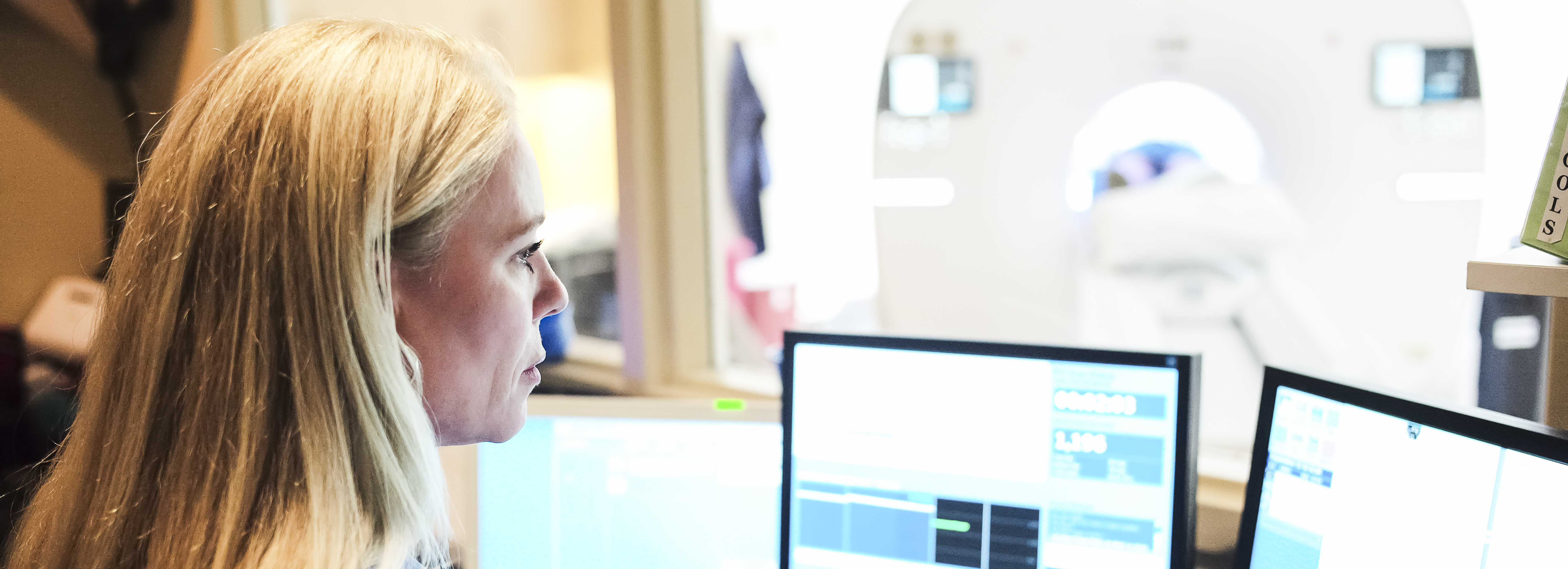
Kristin Petkavich, lead PET/CT technologist at Evergreen Radia Imaging Center, agrees. “Precision DL makes the images significantly clearer, and radiologists are able to see smaller lesions than they have previously been able to.”
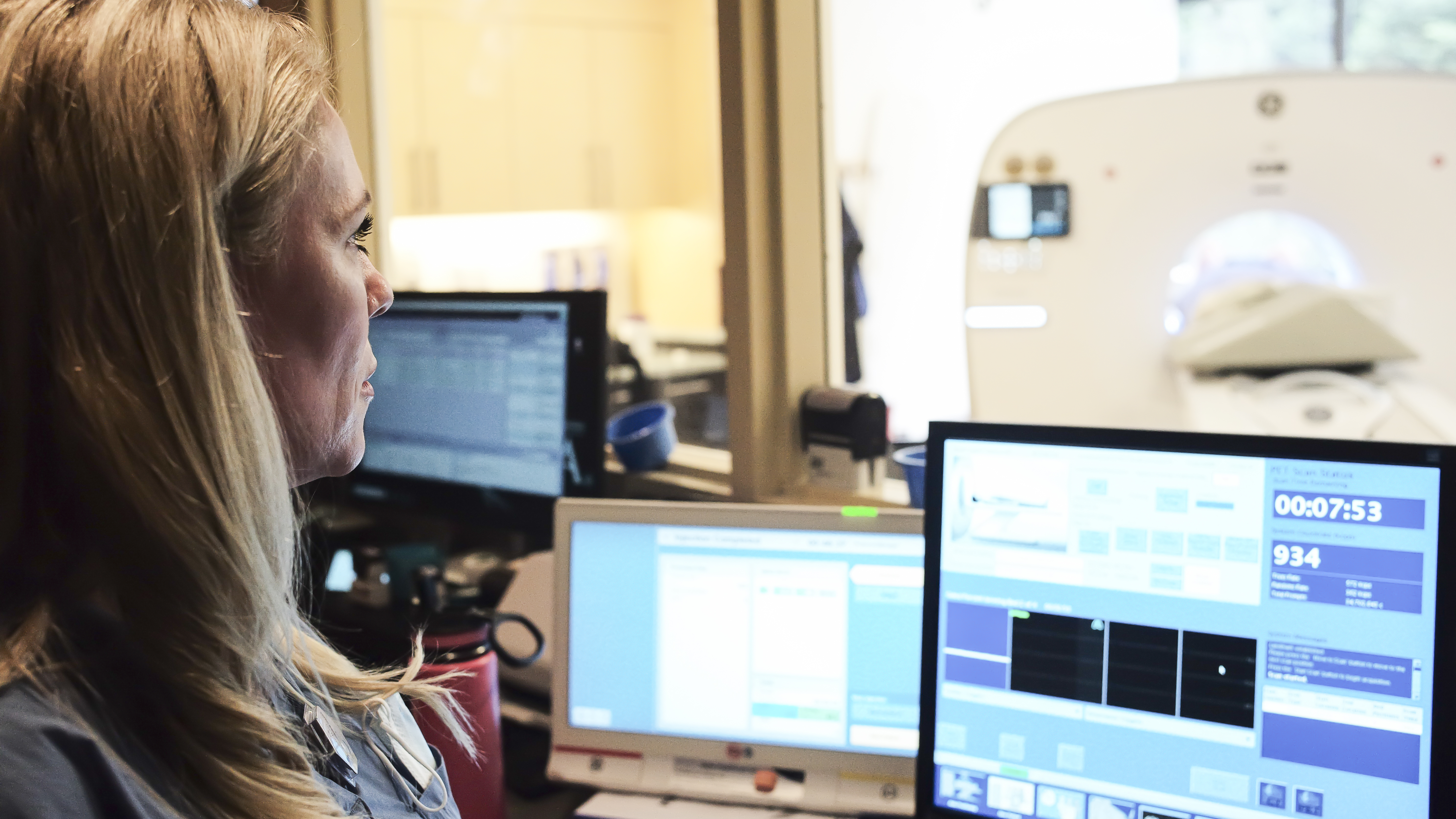
Kristin Petkavich
In addition, while early PET/CT technology required 30 to 40 minutes to complete a full study, the AI-driven process takes just six to eight minutes and the accuracy of the images is not impacted*.
For patients, like Kerri, who was diagnosed with an aggressive form of Stage 3c breast cancer, this means that care teams are able to make assessments of patients' treatment based on the information gathered on the scans, without requiring them to be inside the machine for long periods of time to complete the full study.
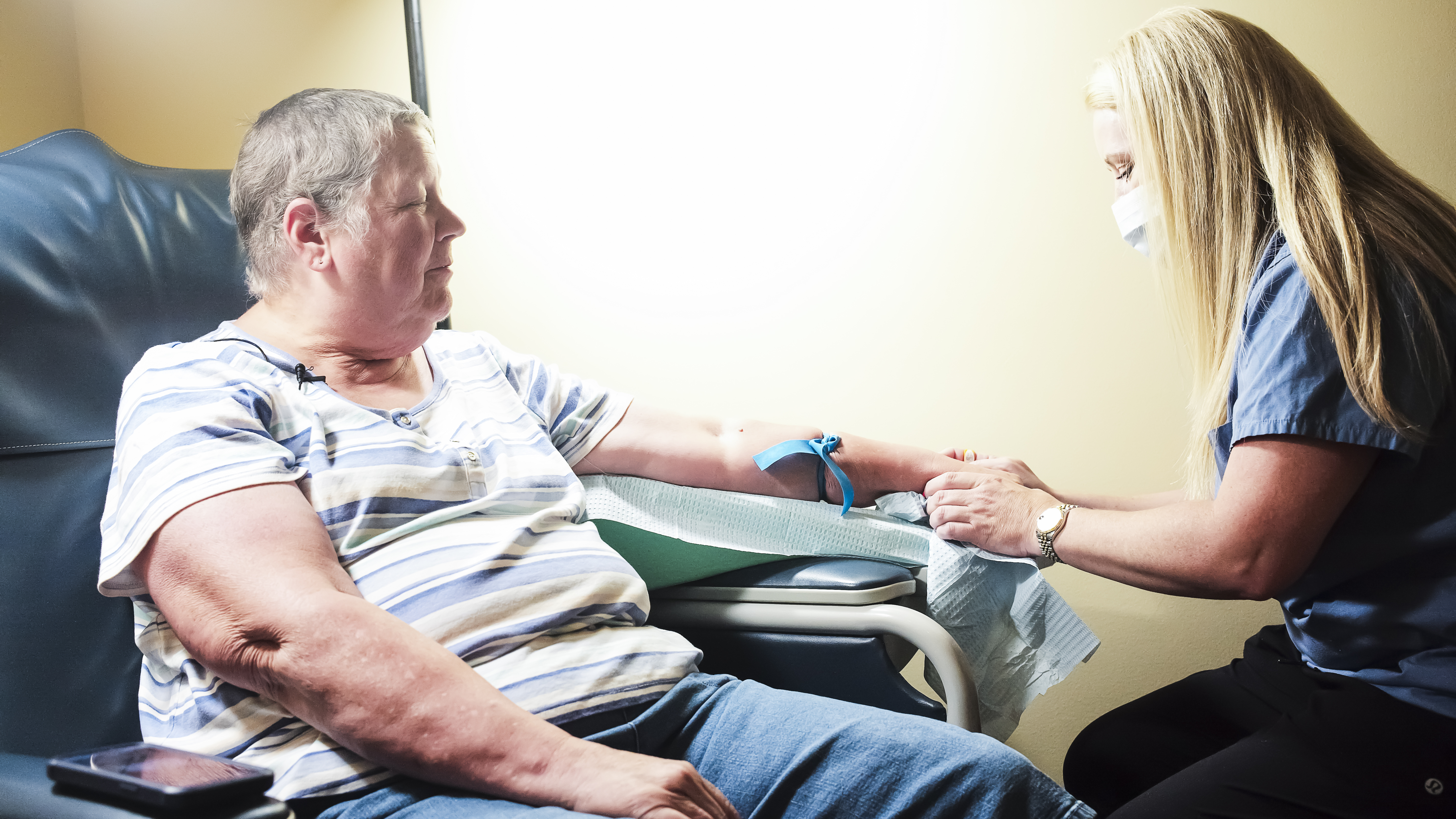
Kerri and Kristin Petkavich
After a mastectomy, chemotherapy, and radiation, “they were hoping they got it all,” Kerri says. But imaging showed that Kerri’s cancer was lingering in her lymph nodes, including two in her neck.
Recently, after the cancer markers in Kerri's blood came down significantly, doctors decided to perform a follow-up scan to establish a new baseline for further treatment. Her follow-up scan confirmed that the two affected lymph nodes in her neck had responded to treatment.
“With the current level of technology with Precision DL and PET imaging, we’re able to provide very rapid, very accurate results to patients and their doctors,” Dr. Stambaugh says. “We couldn’t have done this five years ago. And I think five years from now, we’ll be doing things that we can’t do now.”
Discover AI’s impact on PET/CT results for Kerri in the video below.
*OMNI Legend 32 cm with Precision DL is comparable to OMNI Legend 32 cm non-ToF reconstruction and can image with 50% of the PET scan time or dose of a premium digital ToF PET/CT system at matched small lesion detectability. As demonstrated using clinical data with an 8 mm diameter liver lesion of known location and 2:1 contrast using a CHO model observer, comparing SNR of OMNI Legend 32 cm with QCHD and PDL to SNR of DMI 25 cm with QCFX.