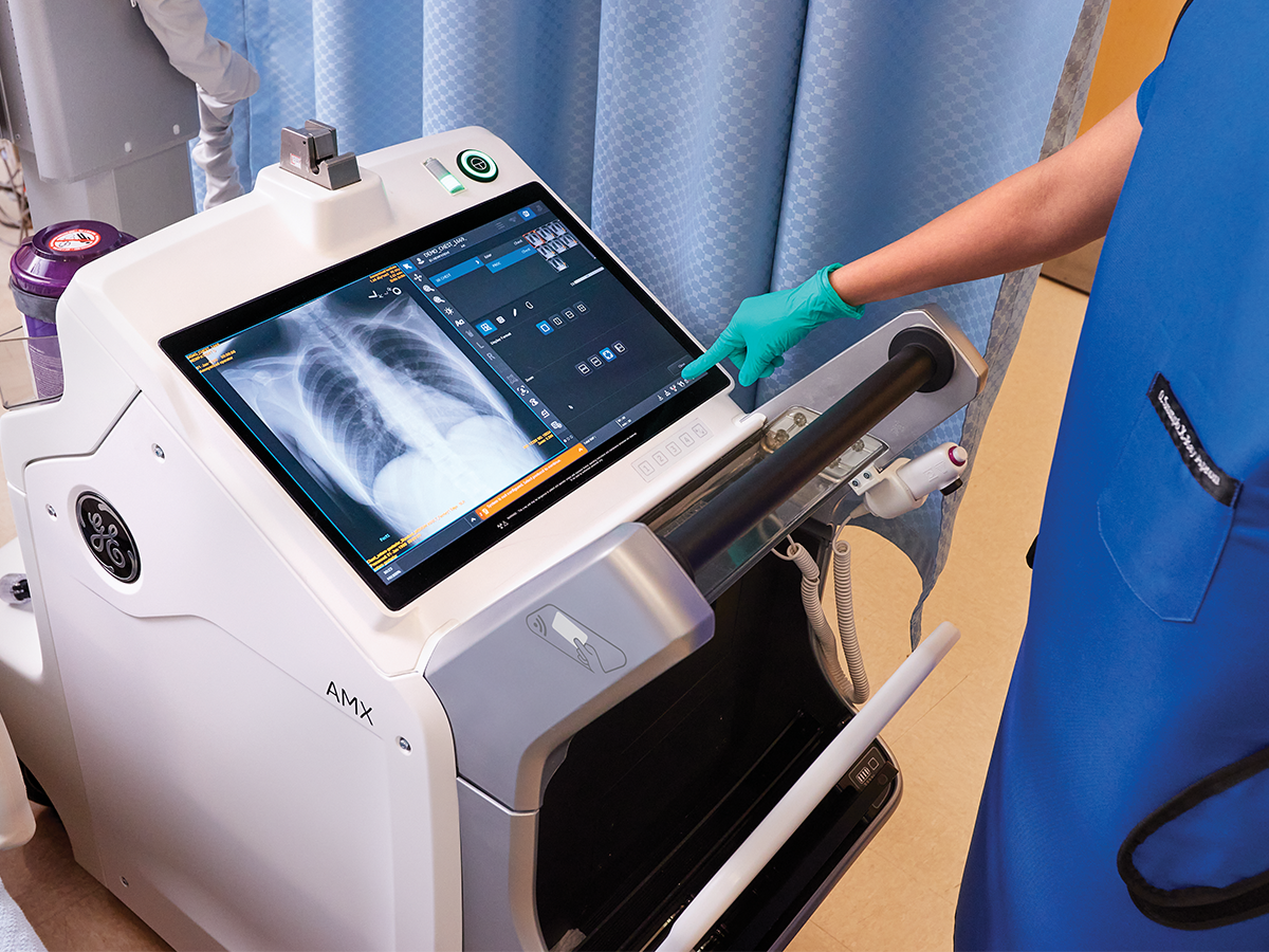
The new frontier of X-ray is developing rapidly, and as such, it’s become increasingly important—and complex—for buying committees to determine precisely what measurements they should use to make decisions on new and upgraded X-ray equipment.
Healthcare facilities know the pivotal role image quality has in enhancing operational efficiency and patient care. That’s why hospitals rank “improving image quality” among their top priorities1 But how do you prioritize what image quality means to your facility?
Facilities with a high volume of chest and abdomen exams have two key considerations: detector size and noise reduction. Determine what detector size will best serve your patients. For example, larger detectors can decrease the risk of anatomy clipping. Smaller detectors may increase clipping risk but are easier to move around an exam room.
Noise reduction and contrast enhancement are also important considerations for large chest and abdomen exam facilities. It's imperative to scrutinize how vendors address noise and contrast in their image processing algorithms. Requesting sample images for evaluation on the facility's Picture Archiving and Communication System (PACS) provides valuable insights into the efficacy of these techniques.
For facilities with a NICU or that handle a high volume of extremity cases, the detector resolution becomes even more important to your needs. Higher resolution detectors can often show finer detail in the small areas where it’s both difficult and important to see as much as possible. Moreover, the management of metal implants is also a critical consideration for these facilities, as they can impact image quality and consistency across images.
How to achieve consistency in image processing and align that process to your requirements can differ from organization to organization. However, it is increasingly important to both radiologists and technologists, especially in the presence of foreign objects and exam variations. Some vendors rely on the performance of their core algorithms while others use AI on top of core image processing. Ultimately, understanding what approach best suits your needs requires examining the outcomes.
Questions to ask prospective vendors about delivery and installation:
- What is the DQE & resolution of the detector?
- What image processing algorithms are available to enhance the images, such as EMI reduction, grid-line suppression, etc.?
- Are your image processing algorithms configurable?
- Is AI used as part of the image processing? How is it used?
- Is there any risk of image artifacts introduced by AI?
- Can the algorithms using AI be configured to achieve the desired image look?
- What is the consistency of the image processing, and how are you measuring it?
- Does the image look change when there are changes in dose, changes in positioning, or when metal objects are present?
- How are metal artifacts mitigated in the images when metal is present?
- Can I see sample images of the following exams?
1. Extremities with and without metal implants
2. Chest exams of different patient habitus
3. Variations in positioning and techniques
Resources:
1. Source: IMV 2019 X-ray/DR/CR Market Outlook Report
© 2019 IMV, part of the Science and Medicine Group

