For more than two decades, the impact of combining single-photon emission computed tomography (SPECT) imaging with computed tomography (CT) imaging has propelled the utilization of this powerful hybrid imaging modality across a growing list of clinical applications.
Before that, SPECT imaging had long been appreciated for revealing the functional characteristics of disease, paving the way for metabolic assessment of tumors and quantifying tumor processes. With the addition of multi-slice CT, clinicians also understand a disease’s relative structural data, as well its precise location in the body. Hybrid SPECT/CT imaging is enabling clinicians to provide precision diagnostics for patients, making personalized medicine a reality and making it possible to help improve patient outcomes.
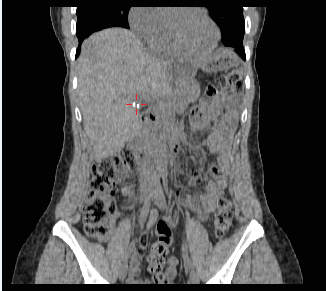
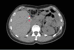
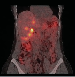
177 Lu Lutathera images (Courtesy of University Hospital of Zurich; Zurich, Switzerland)
SPECT/CT helps enable precision medicine
In contemporary medical imaging, the use of more complex and equally individualized molecular imaging can be critical in optimizing care. Based on the bio-distribution of a radiotracer over time coupled with precision localization, molecular imaging techniques like SPECT/CT map numerous physiological or pathophysiological targeted processes at the whole-body level.
As a proven imaging tool in many clinical areas, hybrid SPECT/CT has rapidly improved diagnostic capabilities because of advances in imaging technologies and the introduction of artificial intelligence (AI)-embedded image processing tools. Hybrid SPECT/CT imaging has even more potential to uphold the promise of precision medicine and personalized care as utilization grows. The discovery and development of novel targeted imaging and therapeutic radioisotopes pairings working with sophisticated detection technology make advanced diagnosis and targeted treatments possible, reinforcing the key role of diagnostic imaging with SPECT/CT in patient management.
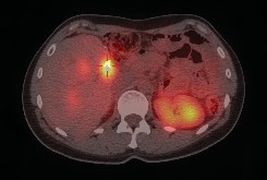
SPECT/CT single-slice (Courtesy of The Miriam Hospital; Providence, RI, USA)
Quantitative SPECT/CT-the new general purpose radiology workhorse
Though there are many clinical applications where planar NM imaging remains the preferred option, the number of applications that support the use of hybrid SPECT/CT imaging for more accurate and comprehensive diagnostic and treatment monitoring information is growing. In fact, interest in capital equipment planning for SPECT/CT imagers has more than tripled since 2008, and the number of installed SPECT/CT imaging units has doubled since the 2011 IMV Nuclear Medicine Outlook Report was published.[1]
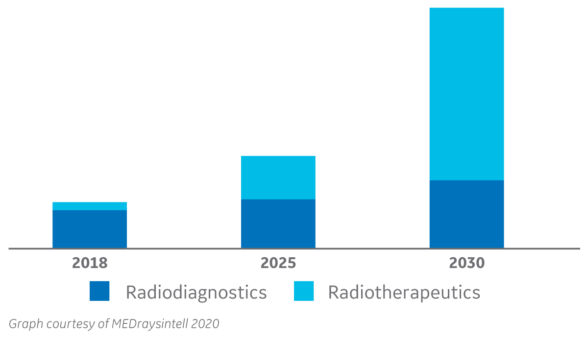
The Global Nuclear Medicine Market, 2018-2030*
13% to 60% |
|
Growth Projection of Radiotherapeutics |
Innovations in SPECT/CT on both the hardware and software side continue to advance the hybrid imaging modality and expand its utility in more clinical areas. For example, StarGuide™ SPECT/CT technology, developed by GE Healthcare, features cadmium zinc telluride (CZT) digital focus detectors, a shape adaptive gantry and an intuitive workflow that automates patient positioning and makes multi-field-of-view quantitative SPECT/CT achievable in routine clinical practice. Hybrid SPECT/CT is currently being used across oncology, cardiology, and neurology. It is used frequently for investigating perfusion and cardiac ischemia, as well as infections, and to image parathyroid disease and thyroid cancer, neuroendocrine neoplasms, bone metastases and prostate cancer.[2]
Voice of customers
“…referral physicians are requesting SPECT/CT and our competitor hospital is getting one, therefore, we expect to lose business unless we can offer it….”
“[hybrid will deliver] better quality imaging and patient care with accurate cardiac interpretations….”
“…building our hospital’s cancer practice and the ability to perform specialized studies like Octreotide is important….”
The shift to digital technology in medical imaging has changed the practice of medicine by exponentially improving the power of imaging diagnostics. With the introduction of solid-state direct-conversion detector technologies such as CZT, innovators continued to push the limits of medical imaging and enabled clinicians to visualize disease with finer detail through high resolution. Hybrid SPECT/CT imaging with these breakthrough digital technologies introduces more imaging data for processing as well as a need for highly sophisticated automated tools and AI-based image processing applications to assist clinicians as they rendered complex diagnoses.
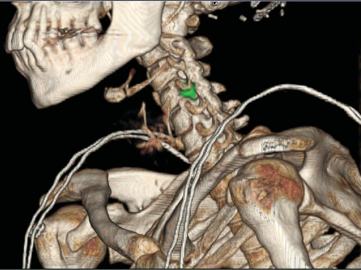
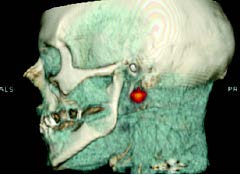
Melanoma on the right auricle (Courtesy of UZA; Antwerp, Belgium, Prof. Stroobants & Dr. Huyghe)
Making the case for continued adoption of SPECT/CT
An increasing number of clinical applications are finding hybrid SPECT/CT imaging is a useful guide to visualizing disease and treatment progress. When there is a high suspicion of active disease or known structural pathology, for example, SPECT/CT may localize multiple sites and define the extent of disease. In cases where a diagnosis has already been established, SPECT/CT is relied upon for treatment planning, including medical, surgical and radiation therapy planning, as well as monitoring patients’ response to a given treatment. The improved diagnostic accuracy resulting from SPECT/CT is also associated with greater diagnostic confidence and better inter-specialty communication.[1] There have also been a growing number of applications for SPECT/CT in cardiac and pulmonary care, such as myocardial perfusion imaging with CT attenuation correction and coronary calcium scoring, and SPECT/CT to evaluate lung perfusion for predicting post-operative forced expiratory volume.
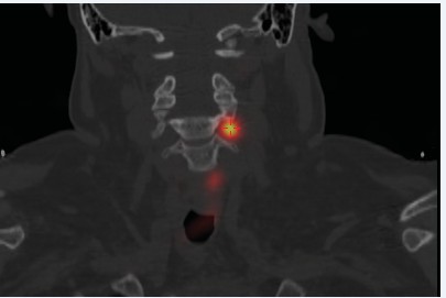
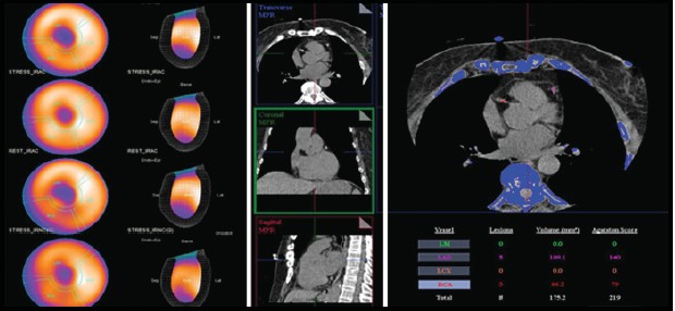
MPI SPECT & CaSc (Courtesy of Rambam Hospital; Haifa, Israel)
The integration of molecular imaging with radionuclide therapy, also known as theranostics, helps increase opportunities for delivering the right treatment at the right time to each patient and helps pave the way towards highly sensitive radionuclide-based precision medicine. The StarGuide™ SPECT/CT system is also capable of scanning in simultaneous dual-isotope mode, so clinicians can visualize multiple tracers with no temporal variation due to the excellent energy resolution of the CZT detector technology. The ability of CZT to improve spatial and energy resolution contribute to the quantitative accuracy of the imaging modality, which together with the incorporation of sophisticated reconstruction algorithms and AI-based processing, is likely influencing the market growth in its adoption.
RELATED CONTENT
- Read about CZT-powered hybrid SPECT/CT imaging
- Learn more about how to Achieve the Promise of Quantitative SPECT/CT in Routine Clinical Practice
- Learn more about GE Healthcare’s SPECT/CT products and solutions
- Download ‘Why Hybrid SPECT/CT?’ overview
- Learn more about our most premium hybrid SPECT/CT camera offering
DISCLAIMER
Not all products or features are available in all geographies. Check with your local GE Healthcare representative for availability in your country.
REFERENCES
[1] IMV 2018 Nuclear Medicine Market Outlook Report.
[2] Israel, O., Pellet, O., Biassoni, L. et al. Two decades of SPECT/CT – the coming of age of a technology: An updated review of literature evidence. Eur J Nucl Med Mol Imaging 46, 1990–2012 (2019). https://doi.org/10.1007/s00259-019-04404-6
*Medraysintell Nuclear Medicine Report & Directory, Edition 2019.

