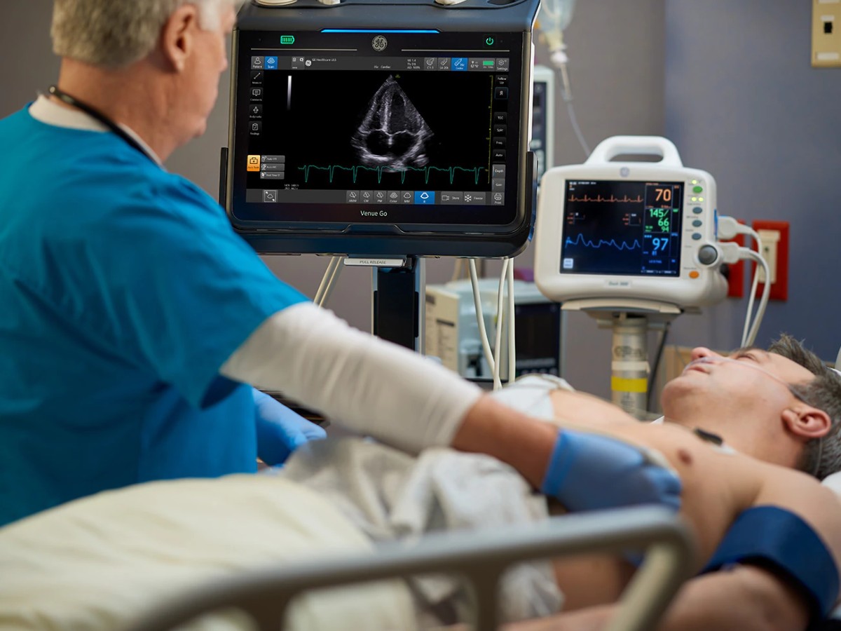
Once an imaging modality tool exclusively used by radiologists, obstetricians, and orthopedists, ultrasound has expanded its application well beyond these specialties. That's thanks to the adoption of Point of Care Ultrasound (POCUS), which offers clinicians the opportunity to perform fast and low-cost scans at the bedside.
POCUS uses have found their way to various care areas as a result of the point-of-care technology's portable, easy to use, low cost, and non-invasive benefits. To support diagnostic decision-making and referrals, cross-discipline teams in emergency care, critical care, musculoskeletal, anesthesia, and pediatrics have implemented POCUS. Moreover, advancements such as artificial intelligence (AI) have helped automate the technology and make it more accessible for these time-strapped physicians.
What are these expanding POCUS possibilities, and how might physicians use them to make more informed decisions? Here's what the literature suggests.
1. Emergency Department Applications
POCUS has a variety of applications in the ED, and is often used as a fast and portable bedside procedure among emergent use cases. Deep vein thrombosis diagnoses have been supported by POCUS with high specificity and sensitivity, for example.1,2 Other applications of this technology in the ED include the detection of left-ventricular ejection fraction (LVEF), wall motion abnormalities, and pericardial fluid.1
It should be noted that some emergent imaging procedures can be complicated and may not be ideal for beginner ultrasound operators. However, the ability of providers to be aware of and detect concerns represents an opportunity for early intervention and prompt referral to specialists or emergency care. Automated tools that rely on AI-based algorithms to inform diagnostics may help overcome the gaps for a more widespread adoption of POCUS.
In addition, while pregnancies are typically assessed and monitored by the obstetrics team, there may be occasions when an ED specialist would need to confirm or date a fetus—including when patients present with potentially related complaints such as nausea, cramping, or missed menses.
Notably, non-OB providers have been able to confirm with 100% accuracy the presence or absence of fetal heart activity in cases of bleeding, and POCUS has additional merit to assess ectopic pregnancy.1 (However, it is preferable to have trained physicians evaluate a suspected ectopic pregnancy given the difficulties visualizing these extrauterine developments.) And if another specialist were to use POCUS at the bedside to assess gestational age, they could likely do so within a margin of three days. Several studies have explored the use of bedside ultrasound to date a pregnancy among general practitioners and emergency physicians alike.1
2. Applications in Critical Care
Physicians across critical care might find particular utility in POCUS for its ability to help detect pneumonia and pneumothorax. As one paper in Current Cardiology Reports notes, the former is often detected through the identification of lung consolidation. For pneumothorax, the detection of lung sliding can help rule out the presence of pneumothorax with a negative predictive value of 100%, while identifying the "lung point" can support a pneumothorax diagnosis with 66% sensitivity and 100% specificity. Another application in critical care is that of detecting ascites, as a sonographic finding showing the presence of abdominal free fluid may support this diagnosis.3
Moreover, POCUS has received increased attention in recent years for its utility with respect to COVID-19 populations in the ICU. As a paper in Journal of Ultrasound in Medicine explores, applications can include monitoring organ function, confirming line placement, assessing thromboembolic disease, and informing ventilator liberation, among others.4
3. Muskoskeletal Applications
Physicians across specialties frequently see broken bones, tears, and other acute injuries. POCUS can be a critical tool for rapidly assessing, diagnosing, and developing care plans for many of these concerns.
Among the indications with the highest sensitivity and specificity for operators are tendon and ligament injuries, such as an Achilles tendon rupture and ankle anterior talofibular ligament strain. Ultrasound may also help indicate the need for radiology referral or CT/X-ray confirmation for other fractures including those of the ribs, tibia, and fibula.1 Rotator cuff tears can also be assessed through POCUS with accuracy rates rivaling MRI.2
4. Anesthesia Applications
Much like that seen with other specialties, POCUS has also been gaining ground among anesthesiologists, who are finding perioperative utility in the tool as they assess patient scans at the bedside when placing nerve blocks, endotracheal tubes, and other interventions.
Recently, a 2020 paper in Current Pain and Headache Reports explored these applications, including that of focused cardiac ultrasound and lung ultrasound, determining their ability to inform diagnostic and interventional care planning before, during, and after surgical procedures.5 Importantly, POCUS has become such an essential part of the anesthesiologist's practice that one paper in Current Opinion in Anaesthesiology posits it could in the future become a part of the mandatory curriculum for anesthesiology residents.6
5. Pediatrics Applications
Many of POCUS' applications in adult populations also carry over to pediatrics, with perhaps the most important one being that of detecting pneumonia, which is the top cause of death among pediatric patients worldwide. For this use, it has shown sensitivity-specificity rates of 96% and 93%, respectively.7 Other applications include the assessment of pulmonary hypertension, free fluid in the abdomen, traumatic brain injury, and POCUS-guided placement of central lines, chest tubes, paracentesis, and other procedures.7,8
Learning POCUS from Colleagues and Training Materials
Despite promising data, as well as indications that interest in POCUS is up among various care areas, there are lingering barriers to implementation. One prevailing challenge is the demand for more training among prospective POCUS examiners, a universal need that has been demonstrated worldwide.1,2,9
Although ultrasound instruction has been incorporated into emergency physician residency training as part of the Accreditation Council for Graduate Medical Education's Next Accreditation System, trainings across other settings and specialties are more difficult to come by.10 In some cases, this challenge has been compounded by a lack of standardized guidelines available to ultrasound operators.9
However, POCUS curriculum is increasingly included in residency programs. Support and advocacy from major organizations including the AAFP, which has endorsed recommended guidelines for family medicine residents, may help this skill set become more mainstream among medical students.11 Manufacturer-provided resources and tools such as GE Healthcare's Venue Learning Labs can also help users master specific equipment platforms through webinars, white papers, and other materials. In addition to manufacturer-provided materials, many industry associations offer POCUS education, including the American College of Radiology and the American Institute of Ultrasound in Medicine.2
When there is an opportunity to learn from other professionals, physicians should take it. Prior literature has affirmed the value of colleague-to-colleague training. In one study, a medical student correctly identified 15 out of 16 abdominal aortic aneurysms with point-of-care ultrasound following three hours of instruction from a vascular sonographer and emergency physician.12
Wherever providers are on their learning journey, they should consider starting small. Depending on the specialty, operators can inform their clinical exams with simple, low-lift exams such as abdominal ascites. They should then gradually work their way up to more advanced applications such as deep vein thrombosis.7 No matter your specialty, AAFP's "POCUS for Beginners" chart can help.11
POCUS: A Powerful Tool
Physicians across many care areas have a significant opportunity to include POCUS in their clinical toolkit. Clinician convenience is a significant advantage, but the benefits of these bedside technologies go far beyond that.
The ability to quickly scan symptomatic patients—and, when possible, diagnose them—promotes improved continuity of care when a radiology referral might otherwise delay findings if patients are required to travel or be transferred. In turn, having this hands-on, “showing while telling” diagnostic tool readily available at the bedside helps foster doctor-patient communication that enables patient satisfaction while enabling providers to bill for revenue-generating CPT codes.11
Still, the utility of point-of-care ultrasound risks can be held back if operators are not suitably trained on the equipment. Learning materials from industry associations and manufacturers can help address this need across specialties in addition to watching exams performed by trained operators.
Ultimately, POCUS offers benefits for clinicians in many settings—and investigations have just scratched the surface of the technology's multidisciplinary possibilities. Physicians and patients alike stand to gain from adding portable sonography to their in-house capabilities.
References:
1. Sorensen B, Hunskaar S. Point-of-care ultrasound in primary care: a systematic review of generalist performed point-of-care ultrasound in unselected populations. The Ultrasound Journal. 2019;11(1). https://theultrasoundjournal.springeropen.com/articles/10.1186/s13089-019-0145-4.
2. Arnold MJ, Jonas CE, Carter RE. Point-of-care ultrasonography. American Family Physician. 2020 Mar 1;101(5):275-285. https://pubmed.ncbi.nlm.nih.gov/32109031/
3. Guevarra K, Greenstein Y. Ultrasonography in the Critical Care Unit. Current Cardiology Reports. 2020;22(11). https://doi.org/10.1007/s11886-020-01393-z.
4. Schrift D, Barron K, Arya R, Choe C. The Use of POCUS to Manage ICU Patients With COVID ‐19. Journal of Ultrasound in Medicine. 2020;40(9):1749-1761. https://doi.org/10.1002/jum.15566.
5. Li L, Yong RJ, Kaye AD, Urman RD. Perioperative Point of Care Ultrasound (POCUS) for Anesthesiologists: an Overview. Current Pain and Headache Reports. 2020;24(5). https://doi.org/10.1007/s11916-020-0847-0.
6. Deshpande R, Ramsingh D. Perioperative point of care ultrasound in ambulatory anesthesia. Current Opinion in Anaesthesiology. 2017;30(6):663-669. 10.1097/aco.0000000000000529.
7. Burton L, Bhargava V, Kong M. Point-of-care ultrasound in the pediatric intensive care unit. Frontiers in Pediatrics. 2022;9. https://doi.org/10.3389/fped.2021.830160 .
8. Singh Y, Tissot C, Fraga MV, et al. International evidence-based guidelines on Point of Care Ultrasound (POCUS) for critically ill neonates and children issued by the POCUS Working Group of the European Society of Paediatric and Neonatal Intensive Care (ESPNIC). Critical Care. 2020;24(1). https://doi.org/10.1186/s13054-020-2787-9.
9. Bashir K, Azad AM, Hereiz A, Bashir MT, Masood M, Elmoheen A. Current use, perceived barriers, and learning preference of point of care ultrasound (POCUS) in the emergency medicine in Qatar – a mixed design. Open Access Emergency Medicine. 2021;Volume 13:177-182. https://www.dovepress.com/current-use-perceived-barriers-and-learning-preference-of-point-of-car-peer-reviewed-fulltext-article-OAEM.
10. Ultrasound guidelines: emergency, point-of-care and clinical ultrasound guidelines in medicine. Annals of Emergency Medicine. 2017;69(5):e27-e54. https://www.annemergmed.com/article/S0196-0644(16)30935-0/fulltext.
11. Shen-Wagner J, Deutchman M. Point-of-care ultrasound: a practical guide for primary care. Family Practice Management. 2020 Nov/Dec;27(6):33-40. https://www.aafp.org/pubs/fpm/issues/2020/1100/p33.html.
12. Mai T, Woo MY, Boles K, et al. Point-of-care ultrasound performed by a medical student compared to physical examination by vascular surgeons in the detection of abdominal aortic aneurysms. Annals of Vascular Surgery. 2018;52:15-21. https://www.annalsofvascularsurgery.com/article/S0890-5096(18)30354-6/fulltext.

