As one of the most powerful diagnostic tools in contemporary clinical medicine, magnetic resonance imaging (MRI) offers clinicians an information-rich opportunity to image and evaluate disease.[1] Clinicians and patients rely on MRI exams for non-invasive diagnostic evaluation in many clinical areas including neurology, cardiology, pulmonology, and much more. The virus that causes COVID-19 infection was designated by the World Health Organization (WHO) as severe acute respiratory syndrome coronavirus 2, where patients generally present with symptoms of respiratory distress, often detected with computed tomography (CT) or X-ray imaging. The COVID-19 pandemic has also impacted the demand for MR imaging, which has been especially helpful in diagnosing COVID-19-related health issues and uncovering ways COVID-19 impacts other parts of the body outside the lungs.[2]
As the pandemic persists, multiple variant strains of the virus are causing COVID-19 infections to surge once again across the globe and increasing stress on our healthcare workers who are tasked with caring for patient communities. With everything from campaigns to promote vaccinations, to the introduction of new MR technology to streamline the daily workload of care providers as they continue to care for COVID-19 patients, leaders in healthcare, government, and industry are doing all they can to provide relief to healthcare workers and ease the daily burden of these healthcare heroes.
Staff burnout due to COVID
Healthcare workers are exhausted. They are retiring. They are resigning.[3] At the beginning of the global pandemic in 2020, the world was vocally grateful to its healthcare providers. For a brief moment in 2021, with the arrival of the vaccines, and downward trends in new cases and deaths, the world breathed a sigh of relief, but it was short-lived. Now, COVID-19 variants Delta and Omicron are proving to be extremely transmissible and causing a global surge in new infections. The US Department of Health and Human Services (HHS) reported COVID-19 hospitalizations are up forty percent in just one month.[4]
With little relief in sight for healthcare workers, leaders in the healthcare industry who see and understand the stress on healthcare staff have resorted to extreme measures to lessen their burden. Hospitals are struggling to hold on to nurses and clinicians, and the influx of patients is never-ending.[5]
New tools to alleviate stress and strain on MR staff
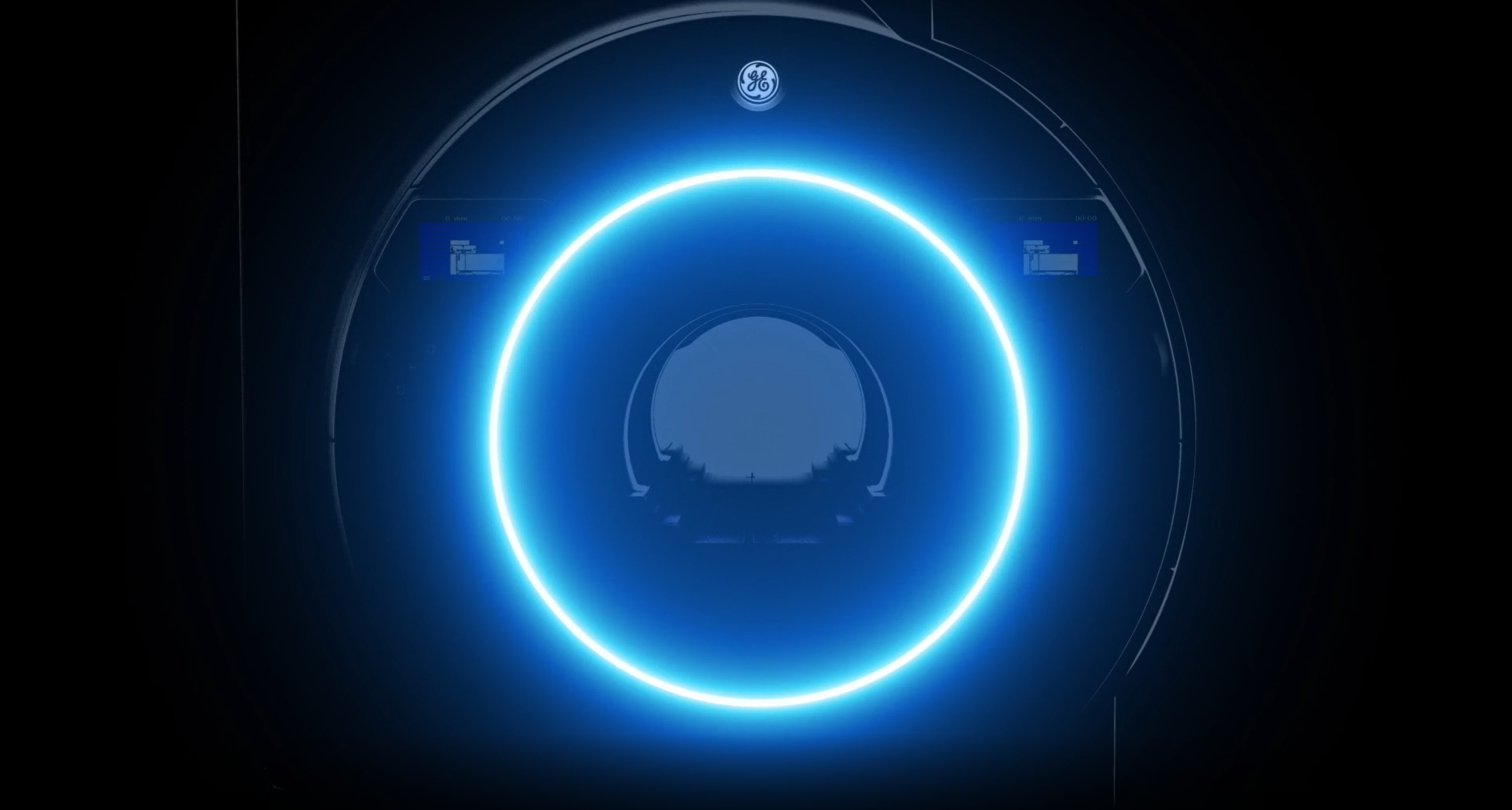
Newly introduced MRI technology from GE Healthcare was designed to alleviate some of the stress and strain on radiology staff who continually care for patients suffering from COVID-19 and other health problems. The aptly named SIGNA™ Hero, a 3 Tesla MRI system, was introduced at the 2021 Annual Meeting of the Radiological Society of North America (RSNA) in Chicago, Illinois.
“The [SIGNA™] Hero was named in honor of all the healthcare workers who continue caring for our global community amidst the COVID-19 pandemic,” said Jie Xue, President and CEO of MRI, GE Healthcare. “Usability is a design imperative for us, and we’re proud to be able to impact the clinical workflow by streamlining the acquisition process with automated positioning tools, prepopulated fields and automated tasks, so we can ease the exam workload of radiology staff.”
The new system was also built to include GE Healthcare’s proven deep-learning embedded technologies that deliver superior image quality to enable accurate diagnoses and reduce exam times for patients.
“The higher image quality enabled by deep learning techniques makes it easier for clinicians to make their diagnoses, so the Hero is as much an operational improvement benefitting radiology staff as it is an achievement in clinical excellence,” said Xue.

Evaluating long-term effects of COVID-19 with imaging technology
Very early on in the pandemic, medical imaging, particularly CT and X-ray were important clinical diagnostic tools in highly suspected COVID-19 cases.[6] And while CT and X-ray continue to be relied upon for COVID-19 diagnosis, the importance of MRI is proving undeniable as clinicians monitor patient recovery and continue to search for the underlying causes of lasting COVID-19 symptoms.
Early reports of long-term health problems associated with COVID-19 infection, referred to as “long COVID” continue to be studied by health experts. Long COVID is described as the syndrome marked by a range of symptoms from breathing difficulties to brain fog that linger after an infection. Some neurologic complications in patients with COVID-19 are commonly seen in hospitalized patients.[7] And more than 80 percent of hospitalized patients may have neurologic symptoms at some point during their disease course.[8]
In a study published in the American Journal of Neuroradiology, a correlation has been found between the severity of the disease in the lungs using CT scans and the severity of effects on patients’ brains, using MRI scans. The results of the study show that by looking at lung CT scans of patients diagnosed with COVID-19, physicians may be able to predict the extent they’ll experience other neurological problems that might be seen on MRI scans, helping improve patient outcomes and identify symptoms for earlier treatment.[9]
COVID-19 variants may have long-term health impacts
The understanding of COVID-19 continues to evolve as vaccinated patients are newly experiencing breakthrough infections, both symptomatic, although less severe, and asymptomatic. While there are many studies tracking disease in symptomatic patients, it has been suspected that infected people who remain asymptomatic play a significant role in the ongoing pandemic, but their relative number and effect have been uncertain.[10]
According to WHO and the US Centers for Disease Control (CDC), the Omicron variant of the COVID-19 virus seems to be more transmissible than other variants of the virus. The CDC expects that anyone with Omicron infection can spread the virus to others, even if they are vaccinated or don't have symptoms.
Until recently, there has not been any formal connection between asymptomatic patients and long-term health effects from COVID-19 infection. Still, there is some evidence building that asymptomatic disease can cause serious harm among some people—including long-term respiratory problems, blood clots, and heart damage, as well as long COVID.
In one recent study, cardiac MRI scans of 1,600 college athletes who had tested positive for COVID-19 revealed evidence of myocarditis, or inflammation of the heart muscle, in 37 people—28 of whom hadn’t had any symptoms.[11] Other studies attempting to evaluate the prevalence of long COVID conditions after asymptomatic infection have varied. From an analysis of healthcare claims, Fair Health, a US healthcare non-profit organization, found that about twenty percent of asymptomatic patients went on to become long COVID patients and still other studies have estimated even higher percentages.[12],[13]
The ability for MRI to identify and track these and other long-term manifestations related to COVID-19 infection is critically important as the body of evidence continues to be collected about the long-term effects of COVID-19 on infection survivors, both symptomatic as well as asymptomatic.
Advancing imaging techniques to reduce staff burden
To adequately monitor the long-term health of these patients, radiology staff will continue to provide care, including longitudinal MRI evaluations of critical organs such as heart, brain and lungs. The advances in MR imaging techniques and technologies continue to lessen the workload burden on staff and improve the clinical assessments of organ function.
Hear more perspectives on the future of imaging from the leaders of GE Healthcare, including the President and CEO of MR, Jie Xue, by tuning into the broadcast from RSNA, available at Innovation.GEHealthcare.com.
To learn more about GE Healthcare’s new 70 cm, 3.0T MRI scanner, SIGNA™ Hero, click here.
To learn more about the deep-learning based MRI reconstruction algorithm to improve image quality, click here.
Not all products or features are available in all geographies. Please check with your local GE Healthcare representative for availability in your country or region.
REFERENCES
[1] van Beek EJR, Kuhl C, Anzai Y, et al. Value of MRI in medicine: More than just another test?. J Magn Reson Imaging. 2019;49(7):e14-e25. doi:10.1002/jmri.26211
[2] https://www.uc.edu/news/articles/2021/03/uc-research-shows-correlation-between-severity-of-covid-19-in-the-lungs-and-brain-using-mri-ct.html
[3] https://mhealthfairview.org/-/media/3C977E375ADA4D3B9B8D7C6AF3C817B1.ashx
[4] https://www.cnn.com/2021/12/16/health/us-coronavirus-thursday/index.html
[5] https://www.cnn.com/2021/12/15/health/covid-traumatized-health-professionals/index.html
[6] Li J, Long X, Wang X, et al. Radiology indispensable for tracking COVID-19. Diagn Interv Imaging. 2021;102(2):69-75. doi:10.1016/j.diii.2020.11.008
[7] https://www.uptodate.com/contents/covid-19-neurologic-complications-and-management-of-neurologic-conditions/abstract/5-12
[8] https://www.uptodate.com/contents/covid-19-neurologic-complications-and-management-of-neurologic-conditions/abstract/12
[9] https://www.uc.edu/news/articles/2021/03/uc-research-shows-correlation-between-severity-of-covid-19-in-the-lungs-and-brain-using-mri-ct.html
[10] https://www.acpjournals.org/doi/10.7326/M20-3012
[11] Daniels CJ, Rajpal S, Greenshields JT, et al. Prevalence of Clinical and Subclinical Myocarditis in Competitive Athletes With Recent SARS-CoV-2 Infection: Results From the Big Ten COVID-19 Cardiac Registry. JAMA Cardiol. 2021;6(9):1078–1087. doi:10.1001/jamacardio.2021.2065
[12] https://s3.amazonaws.com/media2.fairhealth.org/whitepaper/asset/A%20Detailed%20Study%20of%20Patients%20with%20Long-Haul%20COVID--An%20Analysis%20of%20Private%20Healthcare%20Claims--A%20FAIR%20Health%20White%20Paper.pdf
[13] https://www.medrxiv.org/content/10.1101/2021.03.03.21252086v1.full