Around the world, healthcare systems and hospitals are operating under stress and resource constraints while continuing to provide care for their patient communities. Medical imaging is an integral part of providing diagnosis and delivering care. According to the World Health Organization (WHO), an estimated 3.6 billion diagnostic examinations are performed each year globally.[1] The global medical imaging market size is expected to expand at a compound annual growth rate of 5.2 percent from 2021 to 2028, driven by boosts in demand for imaging services because of factors such as the increasing demand for early-stage diagnosis of chronic disease and rising aging demographics.[2]
Advances in imaging, including faster image acquisition, analysis, transfer, and storage, have not only reinforced the central role of radiology on diagnostics, but have also impacted operational efficiencies across disciplines with speed and clinical accuracy. Patient triage, treatment planning and optimization, and real-time image-guided interventions are now integrated into routine clinical practice in most clinical areas.[3]
Increasing imaging efficiencies with X-ray AI across the health system
Healthcare administrators have identified improving efficiencies as a top area of focus in their facilities.[4] Still, hospitals face other challenges such as burned-out employees, cyber-attacks, and wasted resources. Some hospitals and health systems are therefore beginning to utilize highly versatile tools such as mobile X-ray systems embedded with artificial intelligence (AI)-based tools to automate repetitive processes with accuracy—saving time, effort, and money. Healthcare providers and clinicians employing these tools can spend their time on more important tasks, such as working with patients.
The development of more clinically focused AI solutions is on the rise, and radiology is one area where AI can be very effective. The demand for imaging services is increasing, and today, a radiologist can read anywhere from 20 to 100 scans per day, with each scan potentially containing hundreds or even thousands of images.[5] AI algorithms can help to facilitate image acquisitions for technologists, and help automate segmentation of areas of interest for radiologists. Leaders in the industry, such as GE Healthcare are developing innovative imaging AI tools that are bringing more efficiency to not only the radiology department, but across the health system.
“Advances in imaging technology are only one aspect of the solutions we’re bringing to our customers,” says Andrew DeLao, Chief Marketing Officer for Global X-ray at GE Healthcare. “We’re focused on developing holistic solutions that encompass the latest imaging technologies and AI, along with user-centric, sustainable design that can help improve efficiencies across departments.”
Expanding radiology expertise with X-ray AI in the Emergency Department
The Emergency Department (ED) has become one of the major cornerstones of the U.S. healthcare system, and it also plays a major role in other countries, such as the National Health System in the UK. With the number of annual ED visits trending upwards, more patients are turning to health systems’ urgent care facilities more frequently for unscheduled care. And the percentage of those ED visits requiring medical imaging is significant.[6]
Increasing acute healthcare visits represent a large portion of healthcare services globally, with a 22 percent increase in acute care visits over the last ten years in the UK.[7] And in the US, there were an estimated 130 million Emergency Department (ED) visits in 2018 and in 2019; approximately 22 percent of adults aged 18 and over had visited the ED in the past 12 months.[8]
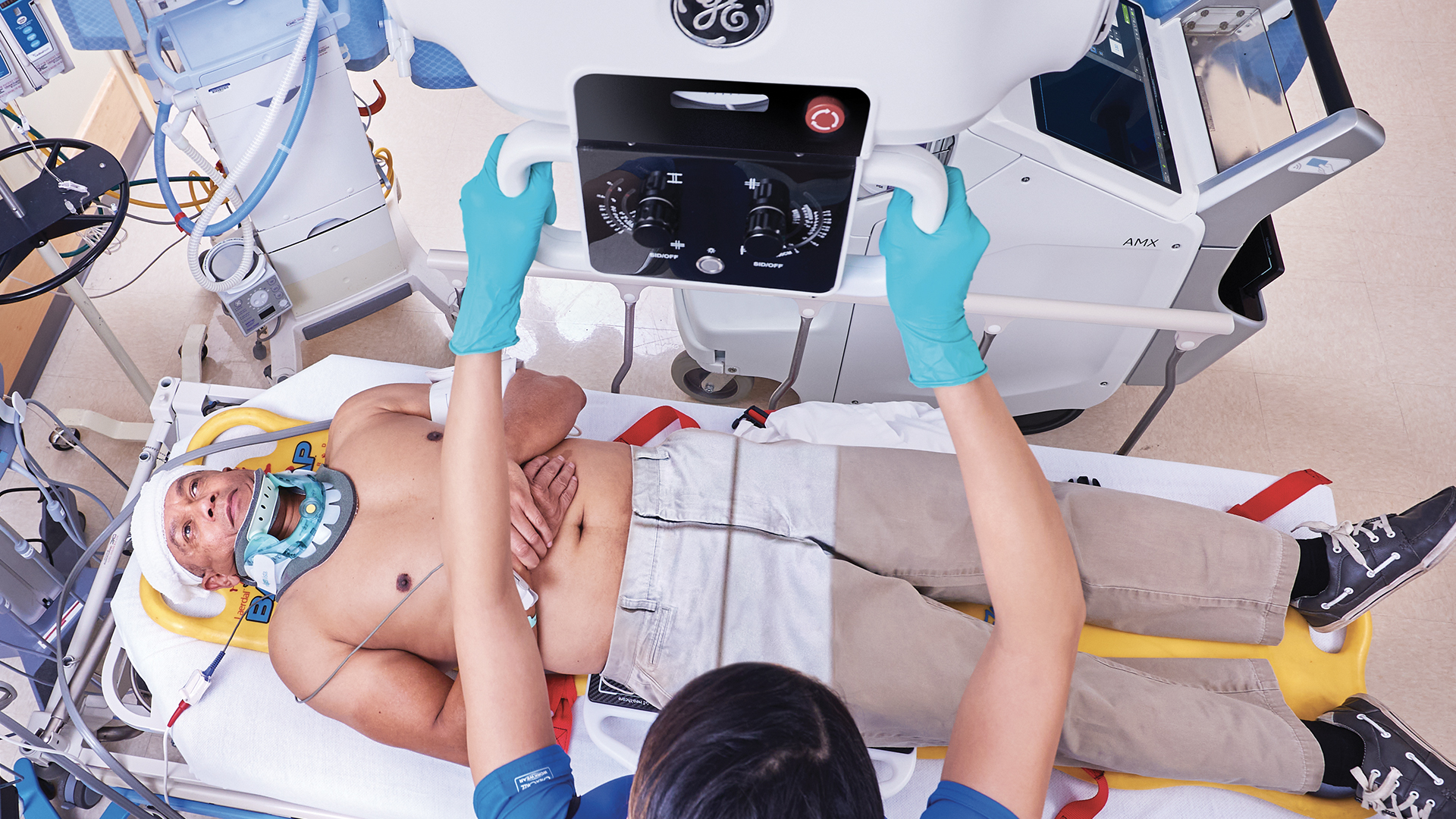
Requests for imaging exams are commonplace in today’s ED. In fact, X-ray is the most requested medical imaging exam, accounting for approximately 35 percent of ED visits that require imaging in the US.[9] Adaptable tools such as mobile X-ray are helping clinicians provide diagnostic care at the bedside, reducing patient transport and saving time.
“There’s a huge scope of unscheduled care dominating the landscape,” said Alex Novak, MD, Consultant in Emergency Medicine and Ambulatory Care at Oxford University Hospitals NHS Foundation Trust. “And there’s a huge shift in cost/benefit tools using imaging diagnostics earlier in the patient pathway, resulting in a great demand on radiology services to deliver on that rapid pathway.”
Dr. Novak is conducting research at Oxford University and works with industry partners GE Healthcare, and the National Consortium of Intelligent Medical Imaging (NCIMI). His current research examines imaging and acute healthcare settings, and more specifically, imaging in the context of changing clinical pathways. He recently presented to radiologists at the British Institute of Radiology to talk about bringing additional insights from radiology tools to the ED physician.
“Why would an ED physician be interested in imaging research?” Dr. Novak asked. “Acute healthcare is a huge burden for radiology. Most of the radiology going on in the ED is actually X-ays, and most of the clinical decisions actually aren’t really seen by a radiologist until a few days later, potentially.”
“That means there’s potential for supportive technologies like AI to really help with the clinical decision-making [in the ED], which is where [diagnostics are] going,” he emphasized.
Streamlining clinical data and insights with imaging AI tools
Innovations in mobile X-ray are currently helping to automate some manual tasks for the radiology technologist and helping to provide reading physicians with clinical decision support. For example, a suite of AI algorithms for X-ray is being used to detect potentially life-threatening conditions on X-ray images in order to help prioritize the most critical cases for reading physicians. Using AI tools in this way can result in improvements in efficiency, quality, and clinical accuracy.
Take, for example, AI’s impact on primary spontaneous pneumothorax—a common medical condition uncovered in the ED. Primary spontaneous pneumothorax is a significant global health problem, with a reported incidence of 18–28/100,000 cases per year for men and 1.2–6/100,000 for women.[10] According to Dr. Novak, patients with this condition often have a normal presentation in the ED. And on an X-ray, in some cases, the pneumothorax might be easily detectable on X-ray, but in others, it's only detected by the eagle-eyes of an experienced radiologist. This provides a gap for AI to step in and help. Dr. Novak explained how that might be possible.
“ED is a pretty common place to pick up spontaneous or traumatic pneumothoraxes, and that’s typically via plain chest X-ay,” Dr. Novak explained. “The images are often variable quality. Sometimes the patients aren’t terribly compliant, [which] makes it difficult to get. And often, we are not really expecting it as a diagnosis, necessarily [because] the patient’s often not very well.”
“[The X-ray] is rarely seen first of all by a radiologist or even a senior clinician like myself. It tends to be seen by the juniors. So, this provides a gap for AI to step in and perhaps support, and to perhaps bridge some of that difference in reporting ability between consultant radiologists and physicians like myself.”
A consulting radiologist remains an important resource for critical cases but radiology AI tools in the ED can help offset the radiologist’s workload and improve the collaborative work between the two departments.
AI tools can also be used for automated measurements, case prioritization, and quality control. These tools are intended to assist technologists to correct anatomical positioning, confirm protocol selections at the time of the scan, and auto rotate images to save time for reading radiologists. To improve triage of urgent cases, the AI tool also automatically analyzes images, upon acquisition, for critical findings such as pneumothorax. Streamlining the workflow, triage notifications are sent directly to PACS and flagged for prioritized radiologist review. Using these tools in the ED, where X-ray is already a mainstay, can bring those radiology insights to the bedside team.
“This is a suite of AI algorithms, which are able to be embedded on an X-ray machine, that can be activated at the bedside and are able to help with workflow prioritization, and detection, automated detection and measurements, and quality control, to a degree,” Dr. Novak continued.
Clinical collaboration to enable imaging insights at the bedside
The ideas shared by Dr. Novak suggest adjusting traditional clinical pathways and finding opportunities to build collaboration between departments—where clinicians can not only share innovative solutions, but impact clinical outcomes. Using the ED as just one example, this refreshed perspective on clinical collaboration can enable imaging insights at the bedside while reducing the burden on radiology.
Transforming acute care delivery with tools that augment the skillset of the ED physician, while creating efficiencies throughout the health system’s clinical workflows and operations supports a more collaborative, sustainable future in a system that is currently under stress.
Learn more about GE Healthcare’s on-device AI algorithm for X-ray, Critical Care Suite.
DISCLAIMER
Not all products or features are available in all geographies. Please check with your local GE Healthcare representative for availability in your country or region.
REFERENCES
[1] https://www.businesswire.com/news/home/20210608005582/en/Global-Medical-Imaging-Market-Report-2021-2026-Analysis-by-X-ray-Ultrasound-MRI-CT-Scan-Nuclear-Imaging---ResearchAndMarkets.com
[2] https://www.grandviewresearch.com/industry-analysis/medical-imaging-systems-market
[3] Rambam Maimonides Med J. 2018 Oct; 9(4): e0034.
Published online 2018 Oct 4. doi: 10.5041/RMMJ.10355
[4] The IMV 2019 Global Imaging Market Outlook Report
[5] https://www.npr.org/sections/alltechconsidered/2017/09/04/547882005/scanning-the-future-radiologists-see-their-jobs-at-risk
[6] https://www.cdc.gov/mmwr/preview/mmwrhtml/mm6222a6.htm#:~:text=The%20percentage%20of%20emergency%20department%20visits%20with%20a%20radiograph%20ordered,Source%3A%20CDC.
[7] Kings Fund Report
[8] https://www.cdc.gov/nchs/products/databriefs/db401.htm
[9] http://www.cdc.gov/nchs/ahcd/ahcd_questionnaires.htm
[10] Melton LJ, 3rd, Hepper NG, Offord KP. Incidence of spontaneous pneumothorax in Olmsted County, Minnesota: 1950 to 1974. Am Rev Respir Dis 1979;120:1379-82.