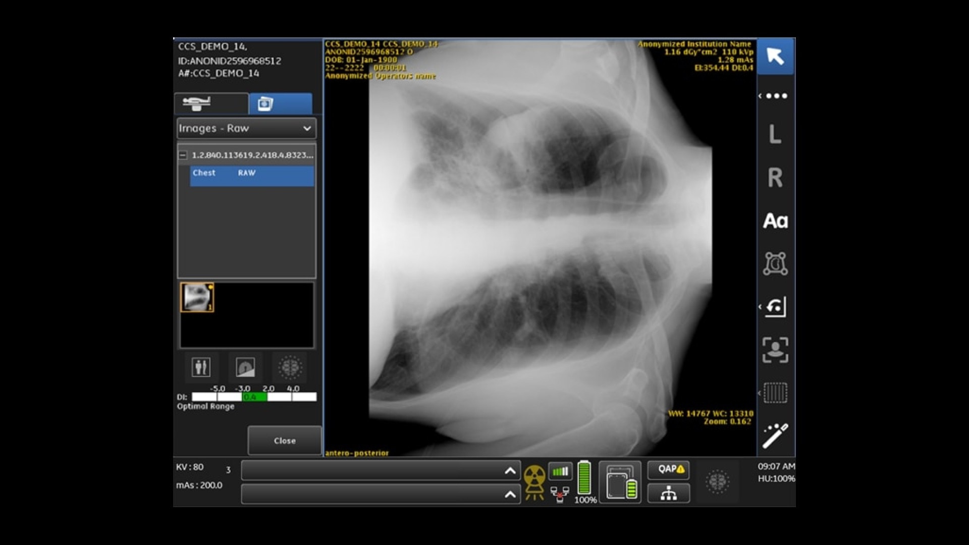
Pneumothorax detection and triage
Given the high number of chest X-rays ordered as “STAT,” i.e., with immediate priority, the triaging of true STATs has become challenging for bedside physicians and radiologists. An average hospital can miss 95 to 352 pneumothoraces each year8-10.
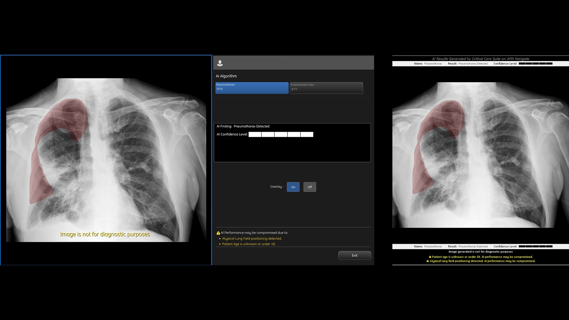
High accuracy11 – Partially localizes 100% of all detected large PTXs and 96.23% of all detected small PTXs.* Limits false alerts (94% specificity), with an Area Under the Curve (AUC) of 0.96.
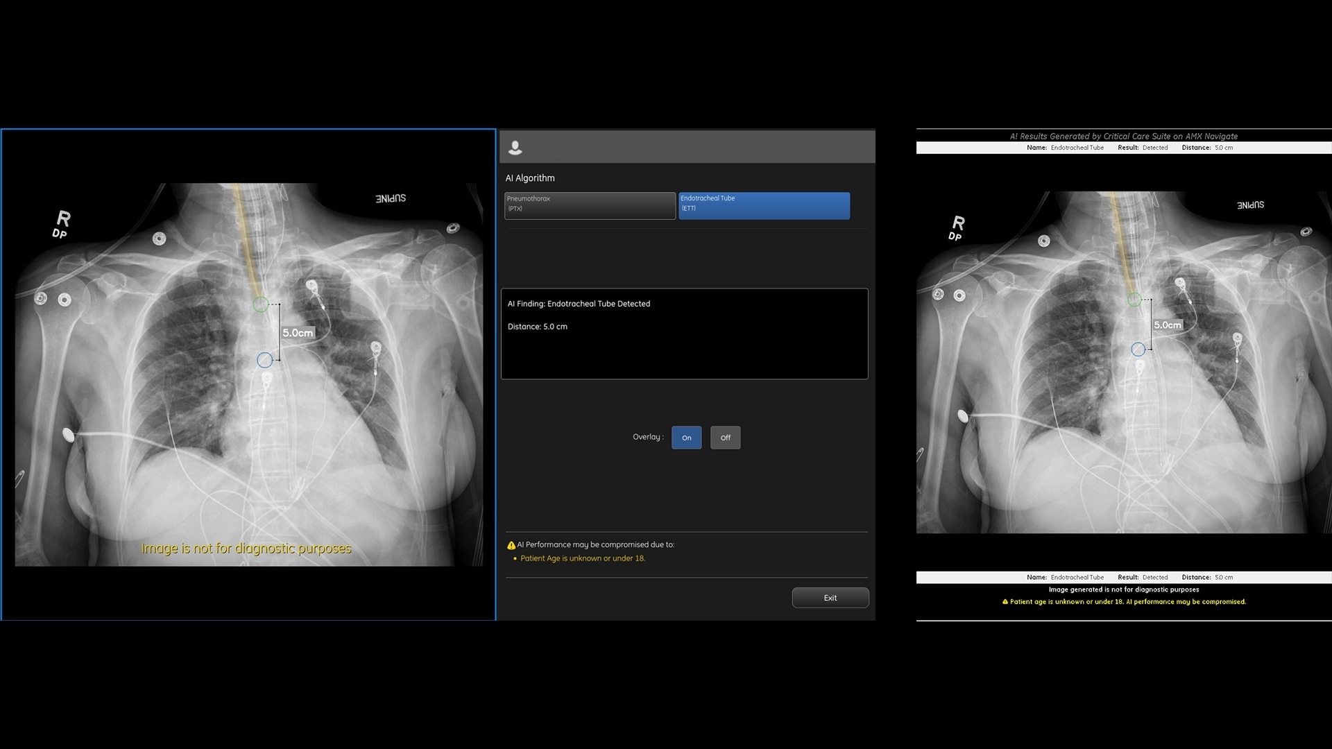
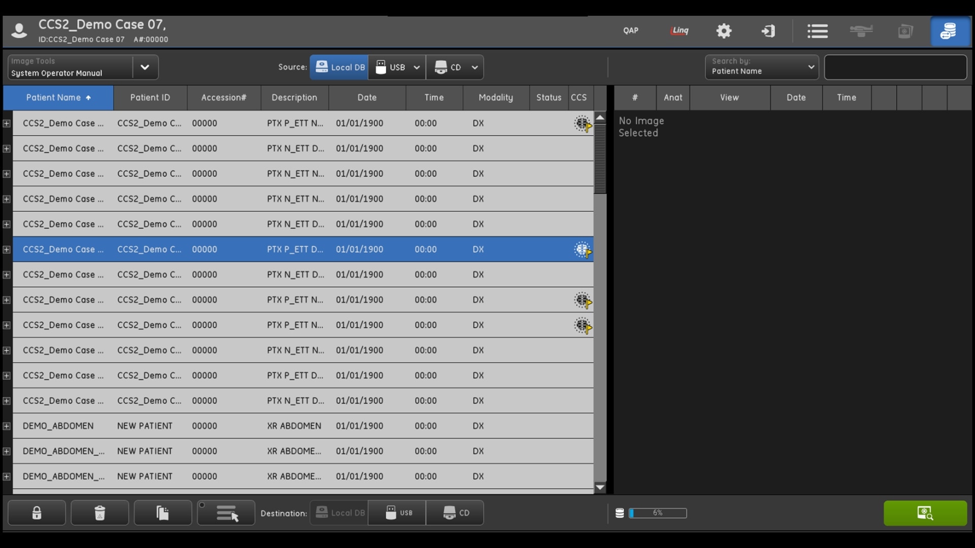
Triage notifications – Sends a secondary capture DICOM image to PACS and presents the AI results to the radiologist. Image flags help enable worklist prioritization and have the potential to expedite review of critical findings.
Overlay display*– When a pneumothorax is detected, an overlay is displayed both on-device and in PACS to assist with localization, along with a graphical representation of the AI algorithm’s confidence level in detection.