Magnetic Resonance Imaging (MRI) is an indispensable tool for imaging and diagnosing a wide range of conditions across all clinical areas, including extremities, cardiac, abdomen, vasculature and oncology. However, medical imaging of certain neurological conditions and disorders remains inconclusive, where often unremarkable imaging results are achieved with current clinical MRI capabilities. These can include mild traumatic brain injuries (mTBI), post-traumatic stress disorder, cognitive disorders, depression, mood disorders, anxiety and panic disorders. Many of these conditions are increasingly recognized as a source of morbidity and impaired brain health and function in healthcare today.[1] Conscious of the critical need for innovation in neuroimaging, industry leaders like GE HealthCare are pushing the boundaries of MRI technology with advanced gradient performance to enable game-changing imaging that can advance the knowledge and understanding of these complex conditions and potentially pave the way for more informed and effective treatments.
“As a leader in medical imaging, GE HealthCare remains steadfast in our commitment to driving innovation in advanced gradient MRI technology,” said Rob Peters, PhD, Global MR Product Manager for Research Systems, GE HealthCare. “Our relentless pursuit of excellence is not only about enhancing image quality but also about transcending the boundaries of what's possible in neuroimaging. With advanced gradient performance, we are poised to unlock new understandings of brain function and structure. This technology holds the potential to revolutionize how clinicians diagnose and treat neurological conditions, ushering in a new era of precision diagnostics in neurology. Our mission is to empower clinical teams with the tools they need to make accurate and informed decisions, ultimately improving the lives of patients worldwide."
Understanding advanced gradient performance MRI
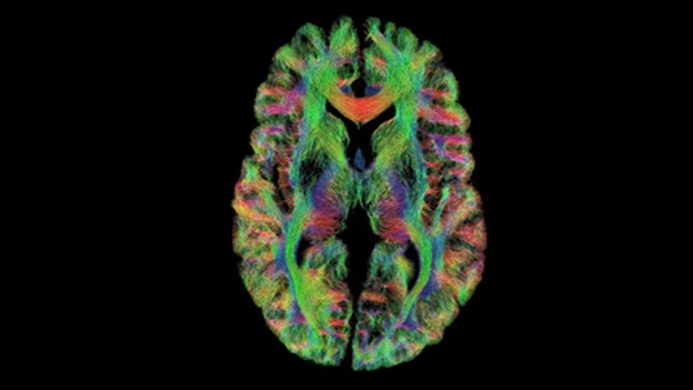
At the core of this pursuit lies the concept of gradient performance. Gradient coils and amplifiers are the components of an MRI system responsible for creating the spatial encoding necessary to generate images. Advanced gradient performance strengthens these capabilities, providing higher resolution and stronger diffusion-weighted contrast in brain images. This breakthrough technology enables radiologists to delve deeper into the intricacies of brain structures and functions, revealing the microscopic behavior of tissues.
One of the remarkable potential applications of advanced gradient performance that is being investigated is the technique of effective axonal diameter mapping.* This technique facilitates the measurement of nerve fiber diameters within the brain, offering insights into the health and integrity of neural pathways. Using advanced gradients, MRI can also illustrate the presence and permeability of membranes and the equilibrium of intracellular-extracellular water.[2] This level of detail has the potential to unravel the mysteries of various neurological conditions, such as mTBI.
Diagnosing traumatic brain injury with clinical MRI
“If I can’t identify the abnormality in the brain, or measure it, then I can’t make it better,” said Colonel Robert Shih, MD, Chief of Neuroradiology and MRI at Walter Reed National Memorial Medical Center (WRNMMC) and Vice Chair and Assistant Professor of Radiology at the Uniformed Services University of the Health Sciences (USU), Bethesda, MD.
In an effort to understand more about diagnosing conditions such as mTBI, several large studies, such as the GE-NFL Head Health Initiative and TRACK-TBI Study (Transforming Research and Clinical Knowledge in TBI) have used advanced diffusion and functional imaging to try to identify mTBI and understand the specific cause of symptoms using 3.0T MR.[3] These efforts have collected important data, but additional research is needed.
Harnessing the power of advanced gradients in neuroimaging research
While several available clinical 3T and 7T MRI systems employ advanced gradient performance for whole-body high-quality clinical imaging, several esteemed institutions are currently conducting pioneering neuroimaging research using specially designed, head-only MRI systems equipped with advanced gradient performance. What if ultra-high performance MR gradients, capable of advanced microstructure imaging, could distinguish patients with mTBI?
GE HealthCare’s (Mesoscale diffusion with Advanced Gradients for Neuro Ultrafast Scanning) MAGNUS gradient platform, installed at Walter Reed National Military Medical Center (WRNMMC) has emerged as a cornerstone of this effort. MAGNUS*, a collaboration between GE HealthCare, GE Global Research Center, and USU, is an advanced gradient MR platform designed to improve diffusion imaging at high b-values without significantly increasing peripheral nerve stimulation or incurring SNR loss.[4] This system is designed specifically for neuroimaging research, allowing for high coil efficiency and excellence in diffusion imaging. It has the potential to unlock new realms of insight into brain-related conditions that have long been elusive to medical professionals.
Seeking a glimpse into the future of neuroimaging
The current strides in neuroimaging research have the potential to transcend the realm of experimental studies and translate into tomorrow’s standard of care. GE HealthCare's commitment to pursuing these advances exemplifies the industry's dedication to realizing this potential. Through the fusion of advanced gradient performance and cutting-edge MRI systems, clinicians and researchers are closer to unraveling the mysteries of the human brain and, subsequently, offering more effective treatments and interventions.
RELATED CONTENT
Join us at RSNA 2023 in the GE HealthCare Innovation theater to tune into: Taking MR neuroscience research & biomarker discovery to the next level with the high-performance MAGNUS gradient system*
DISCLAIMER
*Technology in development that represents ongoing research and development efforts. These technologies are not products and may never become products. Not for sale. Not cleared or approved by the U.S. FDA or any other global regulator for commercial availability.
REFERENCES
[1] https://covid19.nih.gov/covid-19-topics/mental-health
[2] Charles-Edwards EM, deSouza NM. Diffusion-weighted magnetic resonance imaging and its application to cancer. Cancer Imaging. 2006 Sep 13;6(1):135-43. doi: 10.1102/1470-7330.2006.0021. PMID: 17015238; PMCID: PMC1693785.
[3] https://signapulse.gehealthcare.com/the-magnus-gradient-platform-advancing-the-f-7ywwm#id1640718325528
[4] https://signapulse.gehealthcare.com/the-magnus-gradient-platform-advancing-the-f-7ywwm#id1640718325528