All over the world, advances in medical imaging and artificial intelligence (AI) are improving the capabilities of frontline healthcare professionals to diagnose and treat cardiovascular issues. Empowering more healthcare professionals with imaging technology, especially amid shortages of medical sonographers, radiologists, and other healthcare workers, offers a crucial opportunity to improve healthcare outcomes.
Cardiovascular diseases (CVDs) remain the leading cause of death worldwide, claiming an estimated 20.5 million lives globally in 2023. At the same time, 42% of clinicians are actively considering leaving the industry, which underlines a stark disparity: As the burden of treating the diseases grows, there are fewer medical professionals available to treat patients. With cardiac deaths occurring every 1.5 seconds, there is an urgent need for earlier diagnosis and treatment, since more than 80% of these deaths are preventable and premature. Because early detection can significantly improve patient outcomes, the demand for advanced diagnostic imaging—particularly ultrasound, a key tool in identifying CVDs—is rapidly growing.
Because symptoms of CVDs, such as chest pain, can arise suddenly, they trigger millions of emergency room visits annually. In the European Union, for example, 8.6 million patients with diseases of the circulatory system were discharged from hospitals in 2021. In the United States, about 1 million people visit the emergency department each year for acute heart failure, and more than 80% of those patients are hospitalized.
“What’s especially heart-wrenching is that nearly half of all heart failure patients had symptoms for months before they were finally diagnosed. We don’t yet have a holistic cure for the illness, but we do know that adverse events arising from cardiac disease can be prevented if it is caught early,” says Karley Yoder, GE HealthCare’s chief executive officer of comprehensive care ultrasound.
And indeed, frontline healthcare workers agree: “[A common condition] we see in the ER is chest pains, which are concerning for a heart attack and heart failure,” says Sarah Dunphy, a certified nurse practitioner in Atlanta, Georgia, United States. “Anyone could be diagnosed with cardiovascular disease. The more we can learn about it and treat it, the better.”
However, the healthcare labor pool is not growing fast enough to keep pace. For example, in 2021, U.S. clinicians performed nearly 60 million ultrasounds—55% more than a decade earlier—but the number of U.S. sonographers rose only about 44% during that time. In the United Kingdom, there are fewer than 2,000 full-time sonographers, according to the Society of Radiographers, and almost a third of the workforce is close to retirement.
“Every hospital I visit tells the same story: ‘We’re short-staffed, burned out, and there aren’t enough hours in the day,’” explains Yoder. “While technology cannot replace humans, and artificial intelligence can’t clone clinicians, it can hand them back precious minutes.”
Waiting for ultrasounds to be completed by ultrasound technicians and then evaluated by cardiologists can result not only in hospital beds occupied unnecessarily but also delays in patients going home—further straining the healthcare system. The answer to how to diagnose and treat these conditions sooner may lie with enabling first responders with tools and training for cardiac imaging.
Faster scans, fewer bottlenecks, improved healthcare workflows
With approximately 29 million nurses worldwide, and approximately 3 million in the U.S.—as compared with only about 80,000 diagnostic medical sonographers in the U.S. alone—there is a gap for both patient and provider that could be improved with new tools and technology.
Nicolas Poilvert, a machine learning architect at GE HealthCare, was inspired to explore this possibility: “If I could help build software that could make [a] diagnosis a lot easier, more people would discover their condition.”
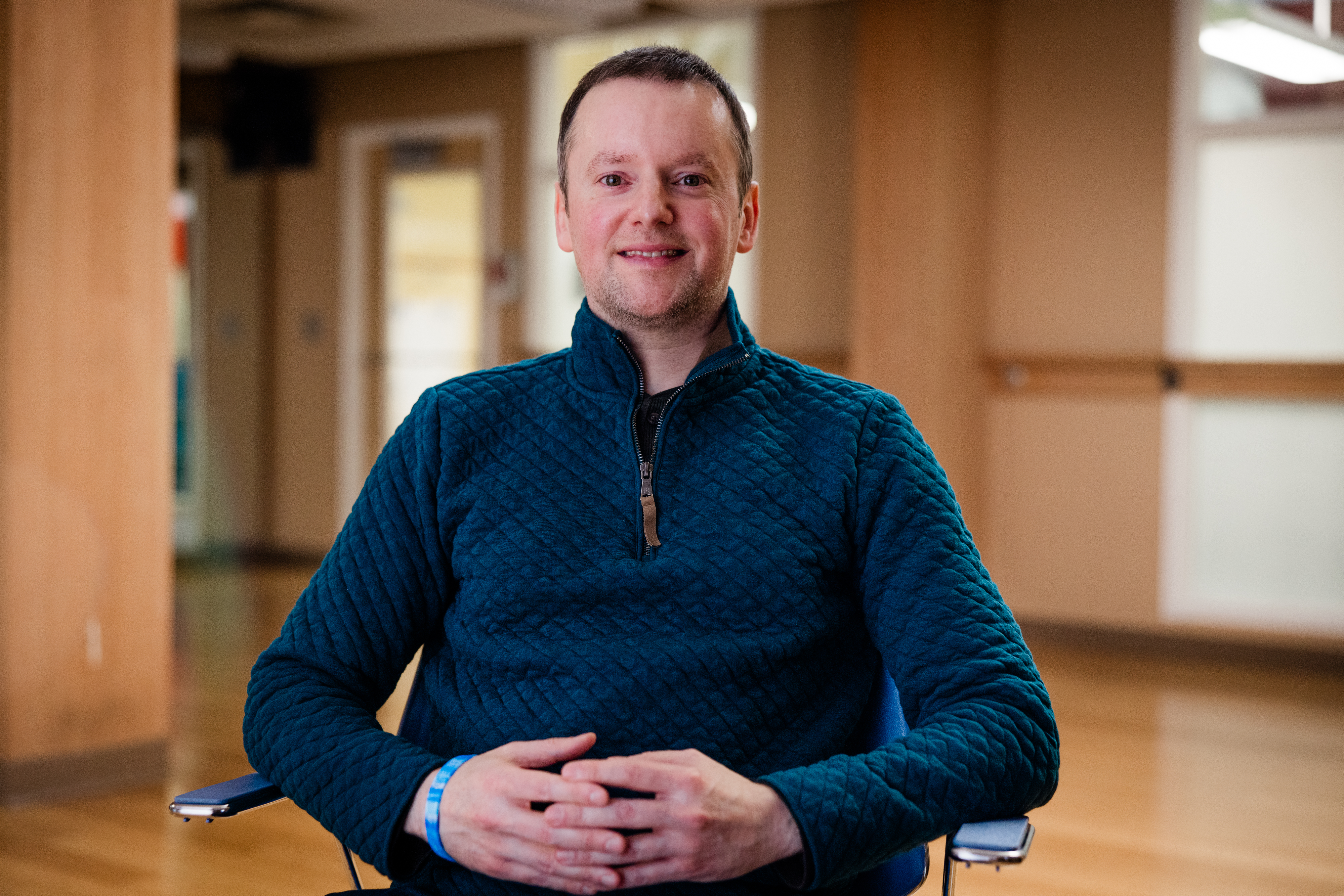
Nicolas Poilvert
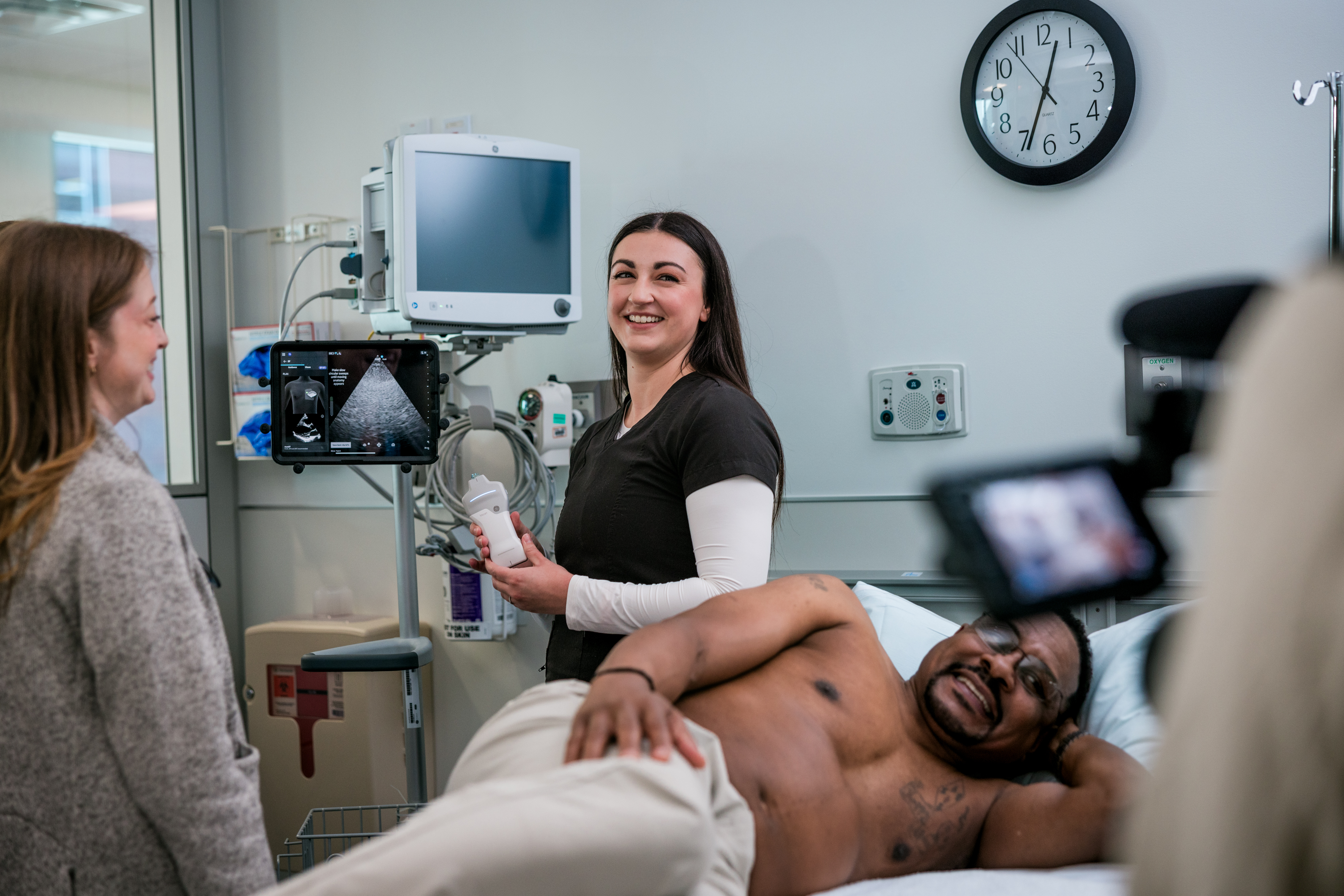
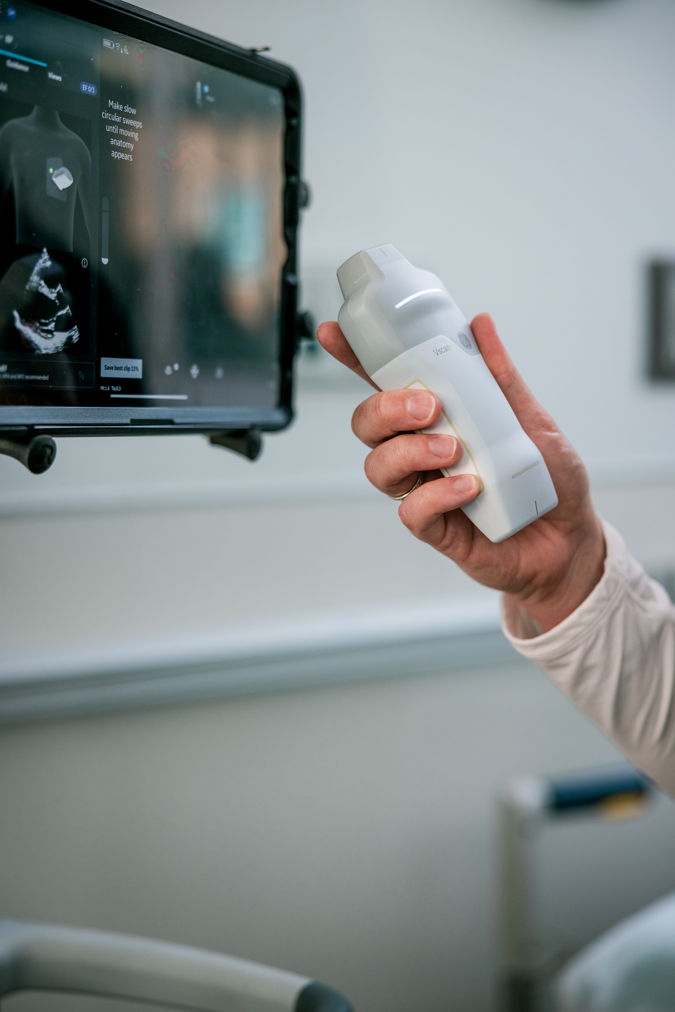
Poilvert contributed to the development of an AI algorithm integrated into a handheld ultrasound device that is designed to support cardiac imaging workflows. The pocket-size device incorporates AI guidance to assist nonspecialists with positioning and disease detection in real time, allowing nurses to perform a basic cardiac assessment at a patient’s bedside.
Widespread adoption of handheld ultrasound technology could help clear the emergency room bottlenecks that delay crucial diagnoses. By routing patients away from overbooked echocardiography labs, healthcare systems can develop more efficient workflows in which specialists can focus on advanced and more complicated cases while frontline workers can be trained and empowered with improved tools to take swifter action to address patients’ urgent needs while also decreasing the strain on the overall healthcare system.
The technology may help deliver results faster. For example, a focused cardiac assessment can be completed with a handheld ultrasound device in less than five minutes, as opposed to 15 to 30 minutes for a traditional echocardiogram—a difference that could be critical during a cardiac emergency.
“AI-enabled devices may offer better and faster images and improved workflows,” explains Eigil Samset, general manager of cardiology solutions at GE HealthCare. “We are building a digital fabric that is connecting everything throughout the patient’s care pathway. It’s not just collecting the images and data, but returning insights that the caregiver can use to understand how the patient is faring and improve access to therapy.”
Enhancing access to improved patient care
From ease of use to cost efficiency, handheld ultrasound technology is already making inroads with a variety of healthcare practitioners, including a growing number of emergency medicine physicians—it is reimagining access to care itself. Such AI-enabled tools are also removing barriers to patient care for healthcare clinics, facilities or systems that could have even more limited resources for acquiring cardiac imaging tools and technologies, such as those in rural or remote communities or those without full echocardiography labs.
Healthcare professionals in rural communities, for example, are increasingly using handheld scanners alongside tele-ultrasound platforms, allowing real-time collaboration with healthcare specialty teams in other locations to ensure more complete and accurate exams and proper next steps to diagnose and treat an individual.
For primary care providers and general practitioners, innovations such as AI-enabled technology can provide an opportunity to respond to patient concerns and possibly eliminate urgent hospital or emergency room visits. “We are already preventing a significant proportion of emergency [room] admissions, which reduces the pressure on our local hospitals,” says Dr. Nabila Laskar,* senior cardiology registrar and research fellow and a specialist in cardiac imaging at one of the largest National Health Service (NHS) trusts in London.
Laskar’s mission is to diagnose and treat heart conditions in her patients long before they become life-threatening. With more tools and technology, there is a possibility for improved patient outcomes. Patients can benefit from quicker diagnosis and decreased wait times—whether in an emergency room or clinical setting. Clinicians benefit from critical information that allows them to make better diagnoses, and hospital systems can reduce strain across the entire healthcare workflow.
“This is what keeps me going: envisioning a future where a woman in a rural town doesn’t drive hours for a pregnancy scan, where an ER nurse could rule out heart failure in minutes, and where a community health worker armed with a pocket-sized probe, AI guidance, and secure cloud collaboration can diagnose early, intervene quickly and save lives,” emphasizes Yoder. “Guided ultrasound, workflow productivity and clinical decision support are more than an imaging framework—they’re a blueprint for equitable care.”
As for healthcare professionals like Dunphy, these new innovations can enhance their confidence in their work and empower them to provide informed care to their patients: “I want to do what I can to help patients feel safe, cared for and heard,” Dunphy says. “Having more information allows me to [do that] quicker and let patients know that we’re here to help them.”
* The statements made here by Dr. Nabila Laskar are based on her own opinions and on results that were achieved in her unique clinical setting.


