CT Perfusion 4D

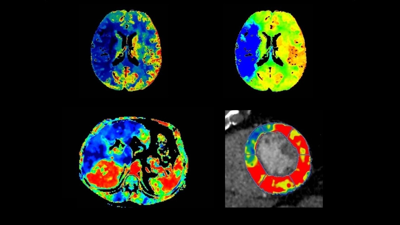
Fast, easy-to-use automated software for analyzing CT Perfusion images related to stroke, tumor angiogenesis and dynamic myocardial perfusion.
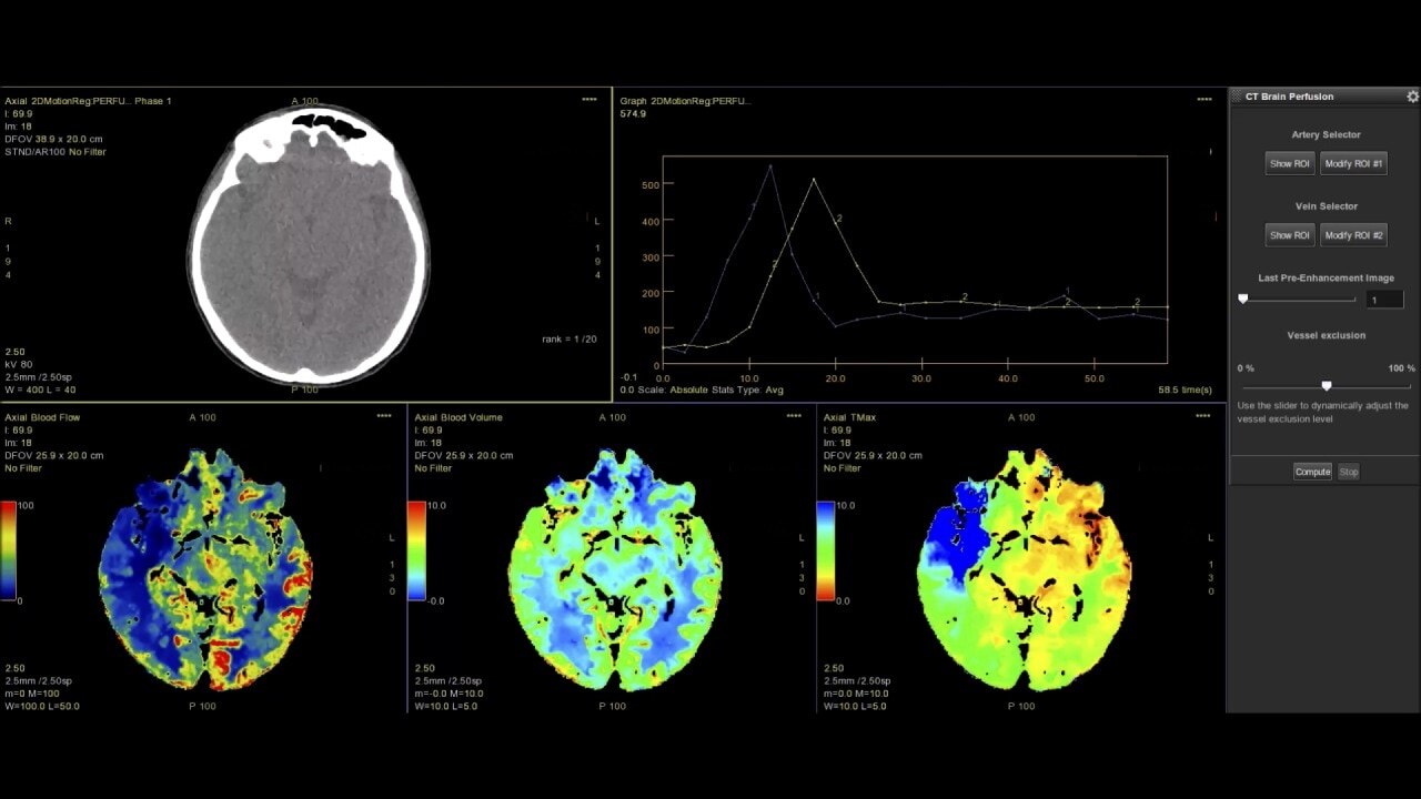
Brain perfusion

Easy-to-use automated workflow for CT stroke and tumor angiogenesis evaluation
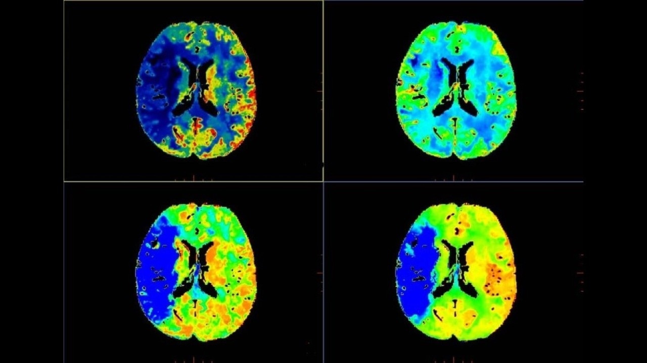
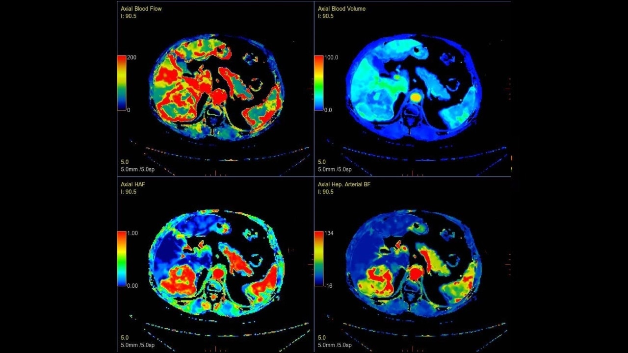
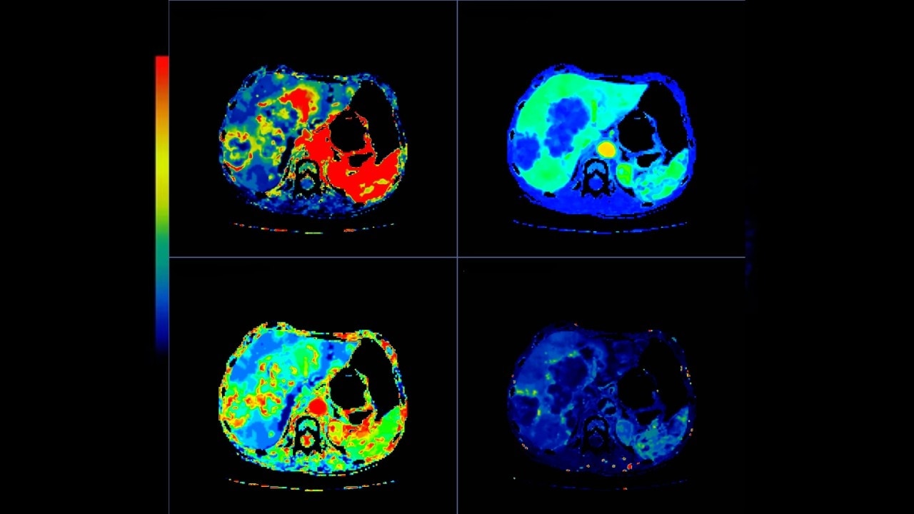
Body perfusion

Optimized protocols for kidney, soft tissue, liver perfusion and other body tumors

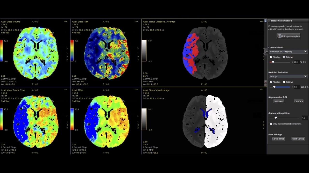
Myocardial perfusion

Complete guided workflow for assessing dynamic CT myocardial perfusion

Brain stroke protocol

  • Image Registration to minimize the effects of patient movement
  • Bone Removal to remove the cranium automatically from the dataset
  • Deep Learning brain ventricle segmentation to prevent ventricular matter inclusion in quantitative results and improve visual inspection of the maps
  • Automated selection of arterial input and venous output. Both can be easily adjusted if needed.
  • Automated generation of all functional maps: Blood Flow, Blood Volume, Mean Transit Time, and Transit Time to IRF Peak (Tmax)
  • Automatically define the symmetry plane to be used for mirroring ROIs and relative thresholds
  • Tissue Classification enables the visualization of regions that are segmented from absolute or relative values, customizable thresholds and user selectable input maps
  • Mismatch volume and ratio are calculated from the modified perfusion volume and low perfusion volume ROIs

CT Body Tumor Protocol

  • Automatically produces functional maps once the user selects an artery using a simple and intuitive workflow
  • Leverages most of the advanced algorithm settings that are part of the Brain Stroke protocol
  • Optimized protocols for pancreas, prostate, kidney, soft tissue, spleen, bone and liver perfusion
  • CT Non-Rigid Registration: Fully automated protocols used for dynamic body acquisitions to register data for use in perfusion analysis

    Dynamic CT Myocardial Perfusion Protocol

    The dynamic myocardial perfusion protocol is laid out in a series of three easy to follow steps. Start by selecting the series that has been generated from the non-rigid registration protocol and then follow the guided workflow:
    • Cardiac Reformat– images are automatically orientated into short and long axis views
    • Segmentation–define the valve plane and the apex of the heart. The software will then calculate the endocardial and epicardial borders and provide a 17 segmented map of the myocardium along with a graphed view for each segment.
    • Functional Maps – the third and final step produces the quantitative data with the color overlay and bullseye maps for blood flow, blood volume, mean slope of increase and mean transit time.
    Get in touch

    Have a question? We would love to hear from you.

    JB29339XX