Cardiac stress testing has become a mainstay for noninvasive assessment of coronary artery disease (CAD), but as evidence emerges about its value across other applications in cardiology, the role of exercise testing stands to expand in the clinic. This is particularly important for heart failure patients, a population that experts predict could increase substantially in the years ahead.
In 2017, for example, the American Heart Association estimated that heart failure diagnoses could jump up 46% by 2030, and by now these numbers may well be underestimated given the projected impacts of both COVID-19 infection and roughly two years of deprioritized cardiovascular prevention.1
As these risks take shape, tools such as stress testing with ECG and imaging may prove to be more important than ever—not only to diagnose and classify the extent of heart failure, but also to identify contraindications to interventions such as cardiac rehabilitation.2
Fortunately, recent developments and evidence in stress testing have helped to inform and evolve the way clinicians evaluate heart failure cases. Here's what's new and why a comprehensive approach that pairs ECG with other assessments such as echocardiography may contribute to a more accurate and efficient cardiac workup.
Adoption of Exercise Testing Across Heart Failure Diagnoses
Exercise intolerance and reduction in functional aerobic capacity are among the key manifestations of heart failure.3 One standard measure of a heart failure patient's functional aerobic capacity or cardiorespiratory fitness is maximum consumption of oxygen (VO2 max), which can be measured with a cardiopulmonary exercise test (CPET).4
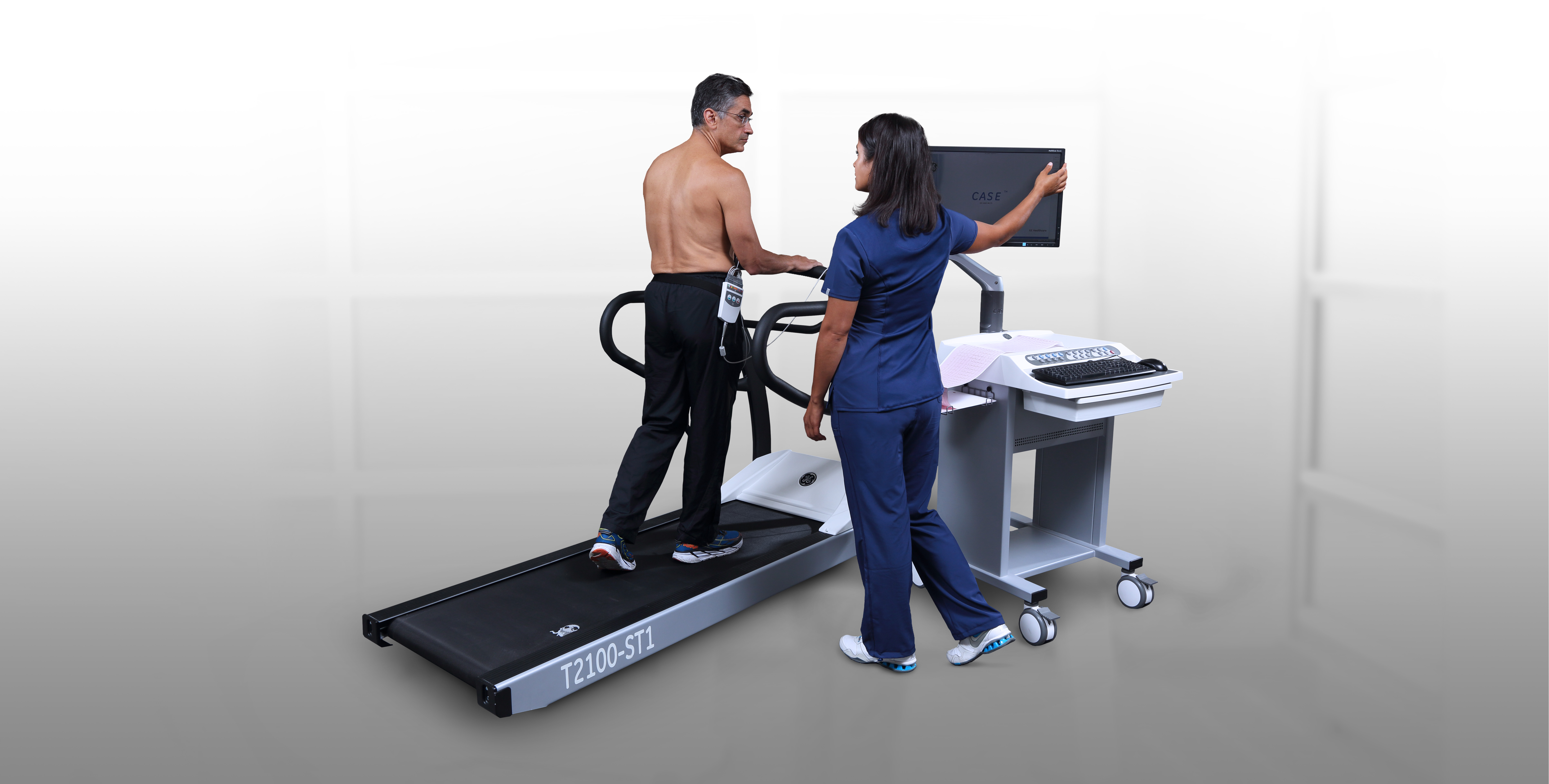
In 2021, the Association for Respiratory Technology and Physiology issued a statement summarizing the utility and best practices for CPET, reinforcing its role as a "gold standard" in cardiorespiratory fitness. The CPET requires specialized equipment that measures the oxygen consumed and carbon dioxide produced during exercise. Standard components of the CPET include a blood pressure cuff, gas analyzer mask, 12-lead ECG, pulse oximeter, and exercise machine.5
Although adoption of CPET has become more universally accepted for advanced heart failure, its documented use in populations experiencing heart failure with preserved ejection fraction (HFpEF) was more limited until just a few years ago. In one 2022 review in Heart, for example, the authors note the potential and promise of CPET for various heart failure diagnoses, including those with normal ejection fraction.6
A New Algorithm for Diagnosing HFpEF
While CPET may not include functional imaging in its configuration, workups that involve stress imaging tests, such as exercise echocardiography, have been recommended for heart failure patients. These assessments can supplement other diagnostic tools, including ECG, and were included in a 2019 consensus recommendation from the European Society of Cardiology (ESC).2
In that guidance, the authors suggest a diagnostic algorithm for HFpEF that originates with a pre-test sequence of standard assessments, such as ECG and echo. Although ECG's diagnostic value here was not determined to be effective in that consensus paper, the authors did emphasize the importance of conducting an ECG to identify Afib, which itself may indicate HFpEF.
If the results of that standard sequence indicate a need for further testing, high-scoring patients revert directly to an HFpEF diagnosis, while intermediate-scoring cases may require a diastolic stress test via bike or treadmill.
As for which is preferred—bike or treadmill—the ESC consensus authors suggest a bicycle test with imaging during the cardiac activity. Otherwise, treadmill exercise can also be conducted with imaging once peak stress has been achieved.7 Notably, one study of severe cases of reduced ejection fraction patients found peak VO2 to be higher on the treadmill.8
Emerging Insights on Heart Rate Recovery
Heart rate recovery (HRR) is the pace at which an exercised heart will stabilize in rate. It has been found to have a predictive bearing on heart failure patients. In particular, recent insights have pointed to earlier HRR measures being more informative and predictive, with one study in the Journal of the American Heart Association finding that a 10-second measure outperformed a 60-second one.
In the study, investigators assessed prognostic values for every 10 seconds within the first minute for more than 40,000 participants. After years of follow-up, the only interval found to maintain its predictive mortality value was the 10-second measure, which supports speculation that parasympathetic reactivation may help to predict poor outcomes.
The authors emphasize the impact that this study could have on future stress assessments, as well as the need to consider short-term recovery rates in using wearables.9
To learn more about the power of the ECG in today's clinical landscape, browse our Diagnostic ECG Clinical Insights Center.
Looking Beyond Coronary Artery Disease
Although the cardiac stress test has a firmly established role in diagnosing and managing CAD, these insights indicate that the technique has wider applications in the cardiology sphere, and even wider beyond advanced heart failure.
As part of a stress test workup, both ECG and functional imaging have important roles to play. Through analysis of heart rate response during and after exercise, the cardiac stress ECG modality stands to complement findings from cardiovascular functional imaging.
New applications across heart failure diagnoses—including in preserved ejection fraction cases—are further validating the utility of stress testing. Moreover, emerging insights on the measurement approach to heart rate recovery could significantly influence how these assessments take place in the years ahead.
In light of these and other developments, one thing is clear: ECG holds considerable and far-reaching value. The time-tested implement is widely available, portable, fast, and inexpensive, and it's a critical tool for cardiologists assessing cardiac activity in heart failure patients with normal or reduced ejection fraction.
References:
- American Heart Association. Heart Failure Projected to Increase Dramatically, According to New Statistics. www.heart.org. https://www.heart.org/en/news/2018/05/01/heart-failure-projected-to-increase-dramatically-according-to-new-statistics. Accessed September 15, 2022.
- Sopek Merkaš I, Slišković AM, Lakušić N. Current Concept in the Diagnosis, Treatment and Rehabilitation of Patients With Congestive Heart Failure. World Journal of Cardiology. 2021;13(7):183-203. doi:10.4330/wjc.v13.i7.183.
- Mayo Clinic. Heart Failure - Symptoms and Causes. MayoClinic.org. https://www.mayoclinic.org/diseases-conditions/heart-failure/symptoms-causes/syc-20373142. Accessed September 15, 2022.
- Mytinger M, Nelson RK, Zuhl M. Exercise Prescription Guidelines for Cardiovascular Disease Patients in the Absence of a Baseline Stress Test. Journal of Cardiovascular Development and Disease. 2020;7(2):15. https://doi.org/10.3390/jcdd7020015.
- Pritchard A, Burns P, Correia J, et al. ARTP Statement on Cardiopulmonary Exercise Testing 2021. BMJ Open Respiratory Research. 2021;8(1):e001121. http://dx.doi.org/10.1136/bmjresp-2021-001121.
- Buber J, Robertson HT. Cardiopulmonary Exercise Testing for Heart Failure: Pathophysiology and Predictive Markers. Heart. Published online April 11, 2022:heartjnl-2021-319617. http://dx.doi.org/10.1136/heartjnl-2021-319617.
- Pieske B, Tschöpe C, de Boer RA, et al. How to diagnose heart failure with preserved ejection fraction: the HFA–PEFF diagnostic algorithm: a consensus recommendation from the Heart Failure Association (HFA) of the European Society of Cardiology (ESC). European Heart Journal. 2019;40(40):3297-3317. https://doi.org/10.1093/eurheartj/ehz641.
- Mazaheri R, Sadeghian M, Nazarieh M, Niederseer D, Schmied C. Performance of Heart Failure Patients with Severely Reduced Ejection Fraction during Cardiopulmonary Exercise Testing on Treadmill and Cycle Ergometer; Similarities and Differences. International Journal of Environmental Research and Public Health. 2021;18(24):12958. https://doi.org/10.3390/ijerph182412958.
- van de Vegte YJ, van der Harst P, Verweij N. Heart Rate Recovery 10 Seconds After Cessation of Exercise Predicts Death. Journal of the American Heart Association. 2018;7(8). https://www.ahajournals.org/doi/10.1161/JAHA.117.008341.