In the ever-advancing field of medical imaging, the quality of clinical images plays a crucial role in providing accurate diagnoses and optimizing patient care. As technology continues to evolve, molecular imaging has emerged as a powerful tool in the arsenal of clinicians. It allows for non-invasive visualization of cellular and molecular processes within the human body, opening new frontiers for disease detection, diagnosis, and patient treatment planning and monitoring.
Evolving image quality in molecular imaging
Over the years, the evolution of image quality in molecular imaging modalities such as positron emission tomography/computed tomography (PET/CT) and single photon emission tomography (SPECT)/CT has addressed some of the historical challenges faced by clinicians to find the right balance between image quality, scan time and dose to the patient. Today, improvements in image quality are addressing these challenges, empowering clinicians with more accurate and detailed information, revolutionizing clinical practice, and profoundly helping impact patient outcomes.
Industry partners such as GE HealthCare are committed to innovating image quality in molecular imaging to inform diagnoses and help improve health outcomes.
"GE HealthCare is dedicated to advancing image quality in molecular imaging, ensuring our customers have access to the finest diagnostic capabilities," said Erez Levy, General Manager of Global Molecular Imaging at GE HealthCare. "By leveraging the exceptional capabilities of our cutting-edge imaging technologies and harnessing the potential of AI-driven image processing, we are evolving image quality in molecular imaging. Our commitment lies in continuing to innovate and deliver enhancements to further improve image quality, empowering clinicians with the intricate details they require to make accurate diagnoses and provide precision care for their patients."
Navigating the historical challenges of image quality in molecular imaging
Historically, image quality has been a significant challenge in molecular imaging. PET/CT and SPECT/CT scanners have revolutionized diagnostic imaging with their ability to visualize physiology within the body and provide functional information about tissues, organs, and lesions. However, image quality can be affected by various factors, including the technical specifications of the imaging device, such as spatial resolution or image sharpness, contrast, and noise.[1] Other factors are contingent upon the patient and organ localization. A patient with a larger build can amplify the impact of scattered photons, and an organ situated deep within the body can be obstructed by overlapping tissues, leading to poor contrast. Any patient or organ movements during a scan can also contribute to a decline in the overall image quality. However, any improvement in one of the areas that impact image quality is usually obtained at the expense or deterioration of another.
The quality of diagnostic imaging has been enhanced by various technological advancements, benefiting both the creation and interpretation of images. Advanced digital detectors, with improved sensitivity and dynamic range, capture more photons emitted by radiotracers, resulting in improved signal-to-noise ratios. This enhancement leads to sharper images with greater clarity, enabling clinicians to discern small details and subtle abnormalities. Molecular imaging technology innovated with digital detectors has made significant progress and has led to improved image quality and enabled the option for a reduction in scan time or patient dose.
GE HealthCare, a pioneer in molecular imaging, innovated its PET/CT detector design to deliver high resolution, as well as increased sensitivity, per the industry standard. This greater sensitivity is an important factor in PET/CT imaging to enable fast scan times as well as the possibility of lowering patient injected dose.
“Improving sensitivity decreases the necessity for the clinician to make compromises that could result in lower image quality or a non-diagnostic scan,” explained Levy. “For example, a pediatric patient is much more radio-sensitive, so the clinician will try to minimize dose, whereas, with an older patient, the clinician may be willing to use a higher dose as they are less radio-sensitive. However due to the level of pain of the patient, they may not be able to lie still for a long exam. Scanners with higher sensitivity offer clinicians the opportunity to maximize image quality, while minimizing scan time or dose.”
In SPECT/CT, system innovations utilizing Cadmium Zinc Telluride (CZT) enable powerful digital detectors that can attain exceptional energy and spatial resolution, striking the right balance of sensitivity and resolution for optimized image quality. By enhancing spatial resolution and reducing noise, modern molecular imaging technologies can provide clearer and more precise images. This is of paramount importance, as it enables clinicians to differentiate between normal and abnormal tissues with greater confidence, enabling improved diagnoses and treatment decisions.
Leveraging AI and deep learning to improve image quality in molecular imaging
Advances in AI and deep learning have proven to be a particularly good fit for diagnostic imaging, primarily for solving a variety of image-classification problems.[2] In molecular imaging, these advances have been successfully applied within image reconstruction and specifically toward the enhancement of image quality.[3] For example, deep learning algorithms can be trained to accurately generate high-quality, full-dose PET images from corresponding noisy, low-dose images.[4]
A leader in AI development across the industry, GE HealthCare created a deep learning-based image processing algorithm that is trained with thousands of PET images created using multiple reconstruction methods, including Time-of-Flight (TOF). Their deep learning software-based tool can take images of already-excellent quality, generated using Omni Legend’s advanced digital detector technology, and further enhance them with image quality performance benefits, such as better contrast-to-noise ratio and contrast recovery, which are most associated with hardware-based TOF.[5]
18F-FDG Omni Legend 32 cm image, reconstructed with Precision DL
Improving image quality across care pathways
Improving molecular imaging image quality can potentially have several benefits for patients across specialties such as cardiology, oncology, and neurology. By enhancing the image quality in these clinical areas, several advantages can be achieved, starting with earlier detection and disease diagnosis.
“We’re committed to providing solutions that enable precision healthcare,” explained Levy. “Precision diagnostics is made possible with image quality improvements that impact small lesion detectability, which is an important factor in clinicians’ ability to detect diseases at their earlier and more treatable stages. GE HealthCare is committed to continuing to invest in innovating detector technologies in PET and SPECT. Additionally, we’re focused on furthering AI and deep learning to keep pushing the limits of image quality.”
In one example, improving the image quality of cardiac images can provide enhanced information about the structure and function of the heart. Image quality improvements can aid cardiologists in accurately diagnosing various cardiovascular conditions, including coronary artery disease, myocardial infarction, heart failure, and cardiac tumors. The advances make enhanced visualization of small lesions possible, as well as areas of reduced blood flow, subtle abnormalities, and the precise location and extent of cardiovascular disease. It can also help clinicians to calculate risk to determine the most appropriate interventions.

Cardiac 82Rb Omni Legend 32 cm image (top row-stress, bottom row-rest)
Across the oncology care pathway, improving the quality of images can enable the detection of smaller lesions and metastases in the early stages of cancer. Advanced molecular imaging can provide valuable information about the functional characteristics of tumors, such as their receptor expression or metabolic activity. With improved image quality, these characteristics can be better visualized and quantified, enabling more precise treatments, such as in Theranostics and treatment monitoring.

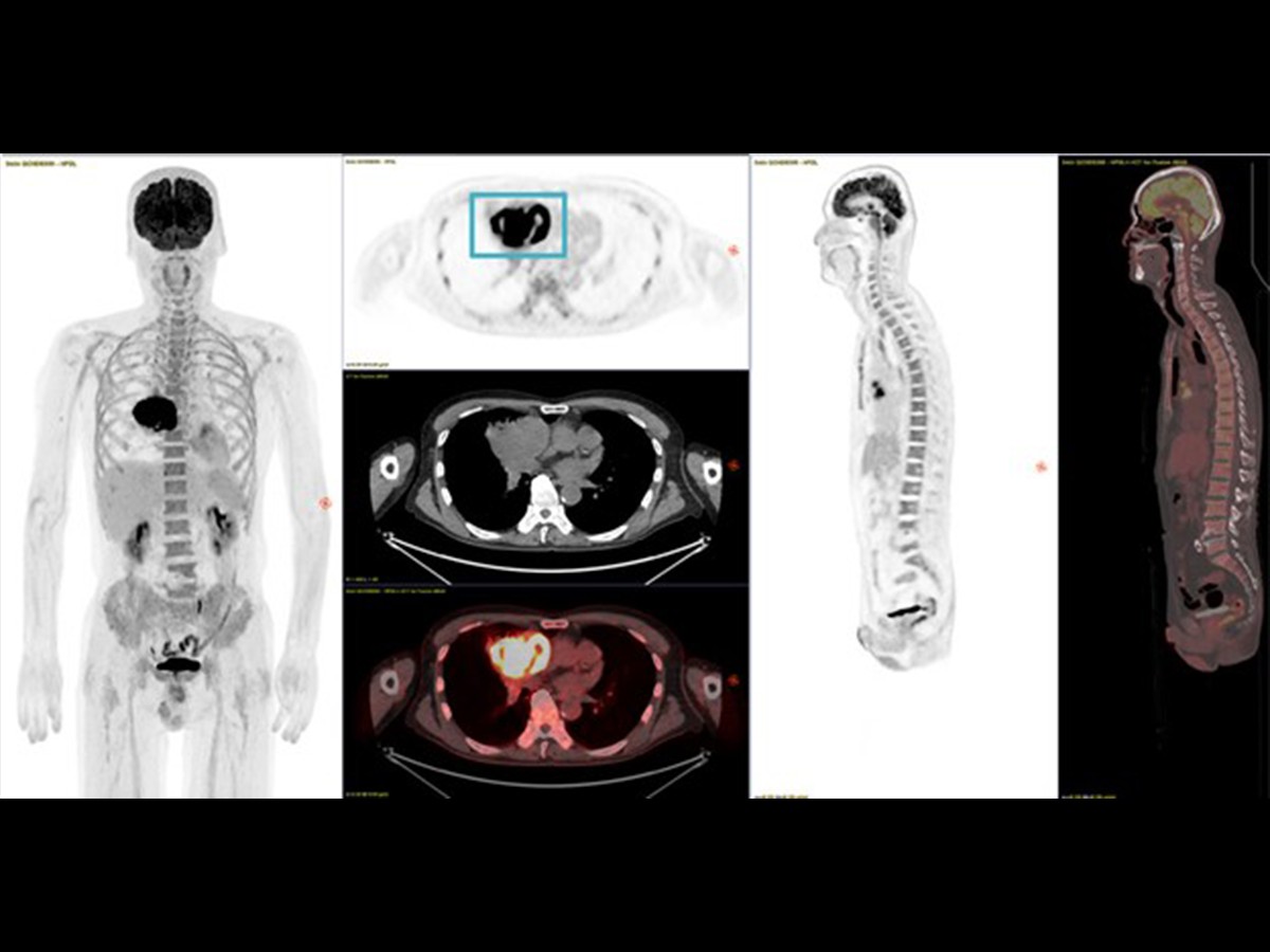
Theranostics imaging, leveraging both digital PET/CT (diagnosis, staging) and digital SPECT/CT (treatment planning and monitoring)
Prostate cancer is one of the most common malignancies affecting men.[6],[7] When treating patients using targeted radiotherapy, or Theranostics, enabled by molecular imaging, clinicians perform targeted prostate-specific membrane antigen (PSMA) PET/CT imaging. This technique allows for highly sensitive and specific detection of prostate cancer lesions. By improving image quality, clinicians can precisely identify the location and extent of the disease, enabling personalized treatment plans.
Enabling the promise of precision healthcare
The clinical relevance of image quality in molecular imaging cannot be overstated. Improved image quality helps enhance diagnostic accuracy, contributing to the ability of clinicians to provide more precise patient care. With improved image quality, clinicians have the potential to identify smaller lesions, detect subtle changes in disease progression, and evaluate treatment response with accuracy and precision. The integration of AI tools and deep learning can aid in improving image quality by quantifying molecular processes, characterizing lesions, assessing treatment responses, and predicting clinical outcomes. The convergence of these technological advancements in image quality is making precision treatments like Theranostics a reality. GE HealthCare is committed to continued innovations in advanced molecular imaging technologies and image quality that help lead to improved clinical outcomes for patients.

RELATED CONTENT
- Learn more about GE HealthCare’s Theranostics solutions
- Learn more about GE HealthCare’s molecular imaging products and solutions
- View the on-demand panel discussion, Theranostics: The future of personalized care and precision medicine

- We invite you to watch our 6-episode series on Theranostics, accepted as one of the most compelling topics in healthcare today. Watch now.
DISCLAIMERS
Not all products or features are available in all geographies. Check with your local GE HealthCare representative for availability in your country.
Clinical images courtesy of 1.) Aizawa Hospital, Japan, 2 and 3.) Rambam Medical Center, Prof. Zohar Keidar, and 4.) Professor Andrei Iagaru, Stanford University
REFERENCES
[1] Vaquero JJ, Kinahan P. Positron Emission Tomography: Current Challenges and Opportunities for Technological Advances in Clinical and Preclinical Imaging Systems. Annu Rev Biomed Eng. 2015;17:385-414. doi: 10.1146/annurev-bioeng-071114-040723. PMID: 26643024; PMCID: PMC5299095.
[2] Hosny A, Parmar C, Quackenbush J, Schwartz LH, Aerts HJWL. Artificial intelligence in radiology. Nat Rev Cancer. 2018 Aug;18(8):500-510. doi: 10.1038/s41568-018-0016-5. PMID: 29777175; PMCID: PMC6268174.
[3] Y. Wang, B. Yu, L. Wang, C. Zu, D.S. Lalush, W. Lin, et al.3D conditional generative adversarial networks for high-quality PET image estimation at low dose Neuroimage, 174 (2018), pp. 550-562
[4] Kaplan S, Zhu YM. Full-Dose PET Image Estimation from Low-Dose PET Image Using Deep Learning: a Pilot Study. J Digit Imaging. 2019 Oct;32(5):773-778. doi: 10.1007/s10278-018-0150-3. PMID: 30402670; PMCID: PMC6737135.
[5] Precision DL with Omni Legend 32 cm data improves Contrast Recovery (CR) by 11% on average and Contrast-to-Noise Ratio (CNR) by average of 23% as compared to non-ToF reconstruction. CR and CNR demonstrated using clinical data with inserted lesions of known size, location, and contrast. Using data from Omni Legend 32 cm, CR and CNR were measured using H-PDL and QCHD.
[6] https://www.who.int/news-room/fact-sheets/detail/cancer
[7] https://www.cancer.org/cancer/types/prostate-cancer/about/key-statistics.html