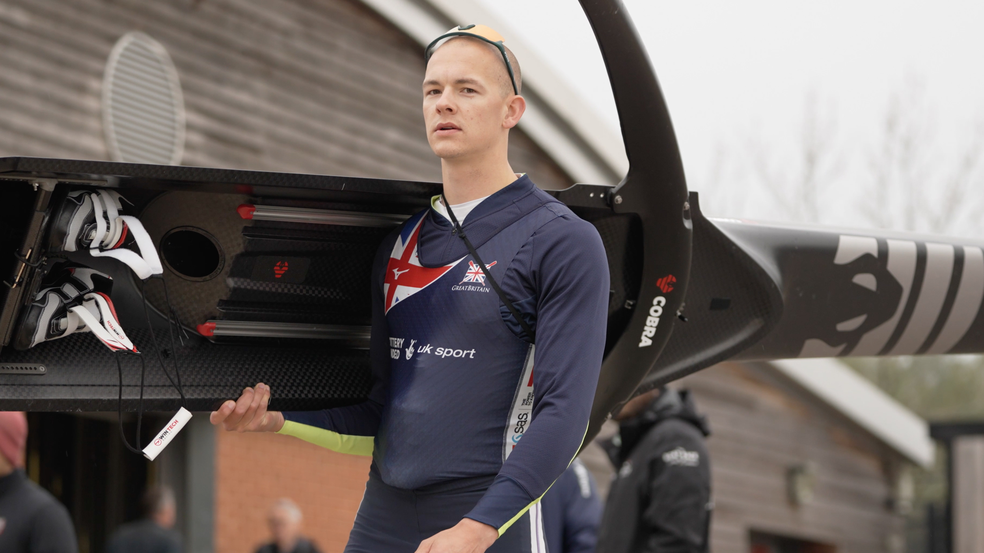
Professional rower George Bourne, of Great Britain, was just weeks away from the Paris Olympic Games trials when he contracted COVID, which has been known to leave devastating heart problems in its wake. Bourne, who won a silver medal in 2022 at the European championships, had symptoms similar to those of teammates who’d developed serious cardiac issues after having the virus. He needed help determining if his heart was up to the continued challenge — or if his rowing career would be put on hold.
“I was worried because I heard that other athletes on the team had real complications,” he says. “Or it could have meant even my health was at risk.”
Getting the right diagnosis
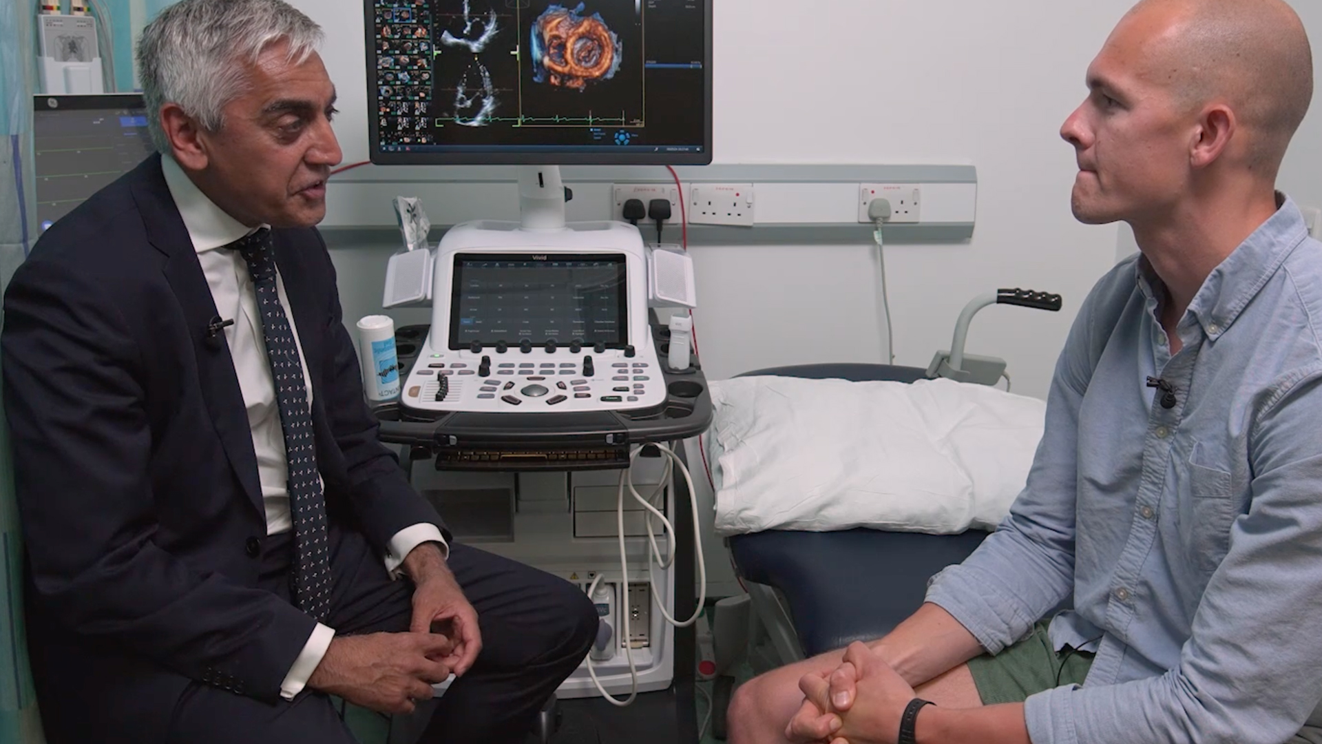
Bourne turned to Dr. Sanjay Sharma, professor of cardiology at St. George’s Hospital, in London, and one of the leading sports cardiologists in the United Kingdom. Dr. Sharma started working with athletes in 1997, after hearing about soccer players suddenly dying on the field from a disorder called hypertrophic cardiomyopathy, an abnormal thickening of the heart muscle. It turns out that sudden cardiac death (SCD) is the leading cause of death among young, competitive athletes. Dr. Sharma began researching how to tell the difference between a heart that was normally enlarged due to the extreme training of elite athletes and hearts that have been damaged by genetics or disease. He now runs the largest sports cardiology unit in the UK.
“It’s very important to make the distinction between physiological cardiac enlargement, at an extreme, or a cardiomyopathy, because an erroneous diagnosis has very serious consequences,” says Dr. Sharma. “It could jeopardize a young life because we’ve allowed someone to carry on competing, or conversely, it can cost an athlete physically, psychologically and financially.”

From left, Dr. Sanjay Sharma and George Bourne
Anyone who exercises intensively for at least four hours a day undergoes a number of structural and functional changes to their heart. Those changes in so-called “athlete’s heart” mean the heart increases in thickness by about 10% to 20% and about 10% in cavity size compared with a normal heart[1]. These changes allow the heart to maintain peak performance for a longer time.
“If you go from rest to exercise, your cardiac output increases,” Dr. Sharma says. “If you’re only doing this for a few minutes, the heart doesn’t have to adapt. But if you’re doing it for hours and hours, the heart must grow.”
While necessary, that growth can mimic cardiomyopathy, especially in male athletes. To determine if he had developed cardiomyopathy or if his heart was safe for exercise after his illness, Bourne underwent a series of investigations, including ECG, baseline echoes, stress echo and MRI scans. Of all the tests, the stress echo, also known as a cardiovascular ultrasound, is the best for diagnosing whether an athlete’s heart is dangerously enlarged[2]. Determining structural defects is important because most sports deaths are due to a structural abnormality.
How technology helps the diagnosis
“Echocardiography is probably the most practical and pragmatic imaging test we’ve got available to us around the world,” Dr. Sharma says. “The most obvious benefits of echocardiography over other modalities, such as an ECG or an exercise stress test, is that it identifies structural heart disease.
“Although an ECG is an invaluable tool for someone like me,” he continues, “as is an exercise stress test in identifying electrical faults and exercise-induced arrhythmias, the only way we can actually diagnose structural heart disease is through an imaging test.”[3]
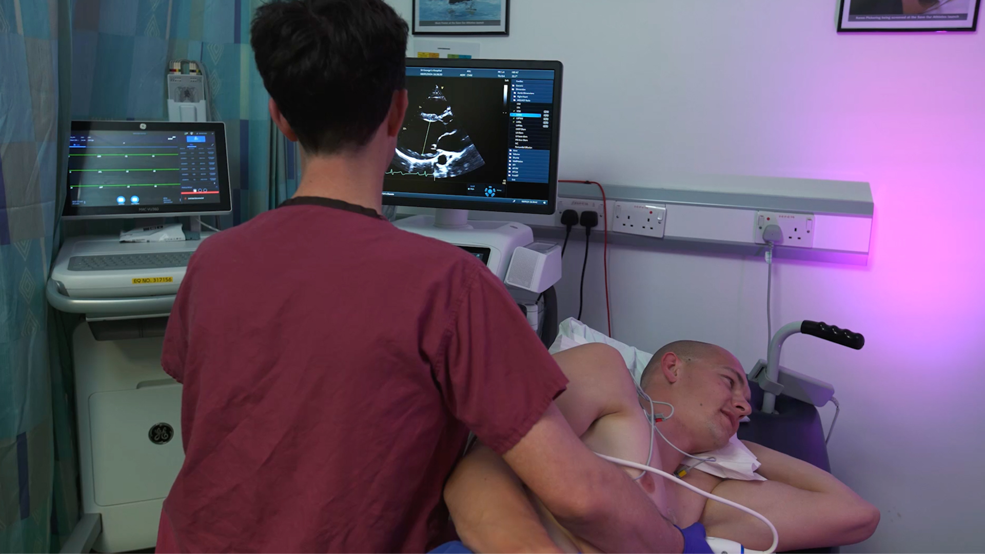
Dr. Sharma performed a baseline scan of Bourne’s heart with GE HealthCare’s Vivid™ E95 Ultrasound System, then while Bourne was exercising he performed additional scans known as “stress echoes,” because they are performed while the heart is working hard. The initial scans ascertain whether an athlete’s heart has physiological changes that are compatible with athletic training. Cardiac ultrasound also allows physicians to assess the condition of the heart’s aorta and even visualize the coronary arteries coming off it.
“In addition to differentiating between ‘athlete’s heart’ and dilated cardiomyopathy, stress echo has a very important role in diagnosing obstructive coronary artery disease [and other cardiac diseases],” Dr. Sharma says.
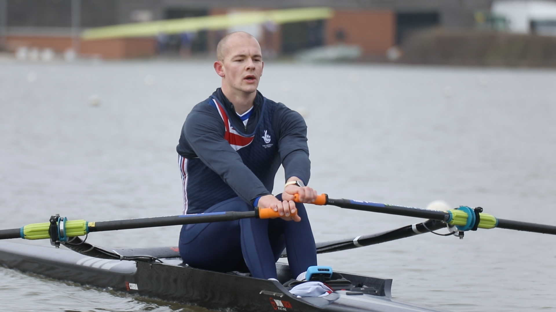
After all the tests, Bourne was finally given the all-clear to resume rowing. “I was relieved,” he says, “but if I hadn’t gotten good news, it’s still a much nicer way to find out here, with the cardiologist, rather than [on the water].”
For Sharma, delivering good news is just part of what he enjoys about collaborating with athletes like Bourne.
“It’s wonderful to work with individuals who push themselves to the limits and demonstrate to us that the human body is capable of so much more than we ever imagined 20 or 30 years ago,” Sharma says. “It’s just great to see that aspiration and desire.”
Dr. Sanjay Sharma is a paid consultant for GE HealthCare and was compensated for his participation in this testimonial/case study. The statements by Dr. Sanjay Sharma described here are based on his own opinions and on results that were achieved in his unique setting. Since there is no “typical” hospital/clinical setting and many variables exist — e.g., hospital size, case mix, staff expertise, etc. — there can be no guarantee that others will achieve the same results.
[1] Understanding the athlete's heart, interview Professor Sanjay Sharma July 2024 – chrome-extension://efaidnbmnnnibpcajpcglclefindmkaj/https://www.gehealthcare.com/-/jssmedia/gehc/us/files/products/ultrasound/vivid-magazine/edition-5/vividmagazineed5profsharmainterviewjb29480xxpdf.pdf – page 7.