Imagine being a sick child, lying in a noisy tube in a strange room without your parents. Every time you move, a voice suddenly filters through, reminding you to stay still. Any movement means you have to start the process all over again — making an already long and strange experience even longer.
For pediatric, geriatric and other patients who struggle to stay motionless, magnetic resonance imaging (MRI) exams can be difficult. MRI is a noninvasive technique that uses powerful magnets — the source of the loud noises — and radio waves to construct detailed pictures of organs and other structures inside the body. MRI is the technology of choice for soft tissues, such as joints, the spine and the brain. It’s also a critical tool for imaging cancer patients, helping clinicians to diagnose and stage the disease, see how patients are responding to therapy, and plan surgery. Traditionally, patients need to spend more than 15 minutes[1] in an MRI machine for radiologists to acquire the best possible images. While shorter MRI scans might send patients on their way faster, they could compromise image quality and, ultimately, diagnostic confidence.
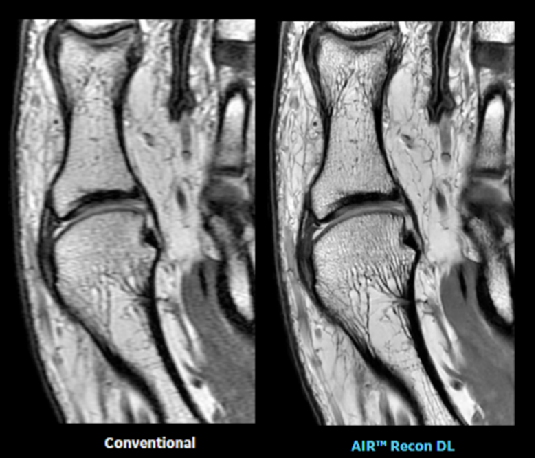
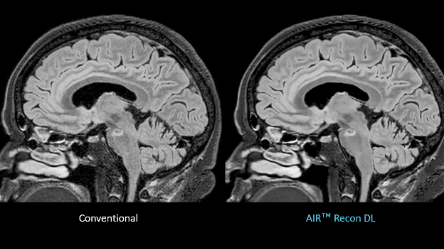
To address this problem, GE Healthcare introduced a new technique called AIR Recon DL in 2020. This pioneering technology uses deep learning to provide sharper, clearer MRI images in as little as half the usual scan time, without compromising image quality. Now the company is announcing that the FDA has cleared software extensions that make AIR Recon DL compatible with additional MRI techniques, including PROPELLER and 3D imaging. By filling in these gaps in clinical coverage, the new software will help physicians to diagnose diseases faster, including those with more complex anatomies.
AIR Recon DL is designed to ease a fundamental trade-off in MRI imaging: scan time versus image quality. A high-quality image — necessary for a confident diagnosis — demands a higher signal-to-noise ratio (SNR) and sharp visibility of body structures. Just as a photographer might leave the camera’s shutter open for a few extra moments in a dark environment to let in more light, radiologists know that the longer the scan, the better the SNR and level of detail. However, that choice comes with trade-offs in both radiology and photography. If you use a slow shutter speed to photograph a basketball game in a dimly lit gym, the fast-moving players will show up as blurs in your pictures. And while patients are not shooting hoops in the MRI scanner, they do breathe; their hearts beat, their intestines contract, and their blood vessels pulse — all of which can distort the resulting image. Longer scans also drag out what can be an uncomfortable experience for the patient, and they limit the number of people an imaging center can serve in a day.
AIR Recon DL combines the best of both options: high-quality images and shorter scan times. Developed on the company’s Edison AI software platform, the deep-learning algorithm reduces noise and improves resolution by operating on raw data acquired during the scan, delivering a clearer final image. In addition, AI techniques like AIR Recon DL can reproduce high SNR and sharpness with less raw data. That means scan times can be shorter, which can reduce the appearance of patient motion, a common challenge for patients and radiologists alike.
The new software release further addresses this issue by making AIR Recon DL compatible with PROPELLER (periodically rotated overlapping parallel lines with enhanced reconstruction), a technique that is insensitive to patient motion.[2] Although PROPELLER delivers high-quality images on its own, it can prolong scan times. When used with AIR Recon DL, PROPELLER will produce the same sharpness and SNR from a quicker scan, improving results for patients who have trouble holding still — such as children and those with neurodegenerative disease — and for people, such as cancer patients, who can’t hold their breath long enough for a traditional high-resolution MRI of the chest or abdomen.
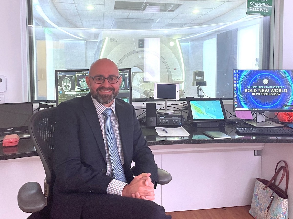
“Patients with chronic liver disease or metastatic disease to the liver are often too sick to be able to perform what is traditionally considered as an adequate breath hold. By acquiring noisier images through a faster acquisition, the breath hold is more doable for the patient, while AIR Recon DL can provide a diagnostic-quality image through de-noising. We would not be able to do this otherwise,” says Dr. Ali Pirasteh (pictured at top), the associate chief of MRI and the clinical director of PET/MRI at the University of Wisconsin School of Medicine and Public Health, who helped assess the new software release.
AIR Recon DL was originally developed to sharpen and clarify 2D scans. Since then, engineers have worked to make it compatible with 3D imaging, which allows for higher resolution than traditional 2D scans and is now a key component to most clinical MR exams to better visualize structures. Radiologists often struggle with resolution in 3D images. Dr. Melany Atkins, medical director of the Fairfax MRI Center and of advanced cardiac imaging at Inova Fairfax Hospital, who helped evaluate the technology for the FDA study, predicts that the new software extension will make 3D imaging faster and clearer. She reviewed AIR Recon DL 3D scans of biliary and pancreatic ducts and was impressed with the results. The images were acquired one minute faster than is common with conventional methods, cutting the typical scan time by about a third.
GE Healthcare is planning to debut the software at a select number of pilot evaluation sites in October and November.
REFERENCES
[1] National Health Service, “How It’s Performed: MRI Scan,” https://www.nhs.uk/conditions/mri-scan/what-happens/.
[2] James G. Pipe, “Motion Correction with PROPELLER MRI: Application to Head Motion and Free-Breathing Cardiac Imaging,” Magnetic Resonance in Medicine 42, no. 5, https://doi.org/10.1002/(SICI)1522-2594(199911)42:5%3C963::AID-MRM17%3E3.0.CO;2-L.