Direct conversion and the changing of the guard for nuclear medicine
Ever since the very first Anger camera was delivered to The Ohio State University Hospital in 1962 for research, nuclear medicine has been on a roll. Over the intervening years, enormous strides have been made in the detection and treatment of cancers and lesions to the benefit of millions of patients.
Sixty years later, the pace and magnitude of those advancements have slowed significantly as the Anger camera has reached its theoretical performance maximums. Yet the overall pace and magnitude of advancements in nuclear imaging have not slowed thanks to the yet-to-be-realized potential found in the Anger camera’s successor: the CZT SPECT/CT camera.
Direct digital conversion
CZT, short for Cadmium Zinc Telluride, is a compound semiconductor material used in the camera’s detector and represents a breakthrough in many aspects of performance over the Sodium Iodide (NaI) scintillator crystals used in the Anger camera. The most notable difference between the two technologies is the fact that CZT crystals directly convert gamma ray photons into digital signals that are able to be read by the detector’s ASIC board. With the Anger camera, the analog signals sent from its NaI crystal require additional and bulky photomultiplier tubes (PMTs) to complete the job.
Our current CZT modules each have 256 pixels as small as 2.46 mm – each capable of capturing events independently via direct digital conversion.
Another prominent difference between the two technologies is pixelization. CZT detector heads from leading manufacturers vary in design, but generally consist of many CZT detector modules. Our current CZT modules each have 256 pixels as small as 2.46 mm – each capable of capturing events independently via direct digital conversion.
Utilizing direct digital conversion, CZT detectors are able to provide more accurate identification of the event location and the energies emitted. The noise and signal loss common to analog NaI-based cameras simply does not come into play with CZT detectors.
Additionally, direct digital conversion can potentially provide:
- Improved image contrast
- Improved multi-isotope imaging
- Faster scanning/lower-dose scanning
- Improved quantitative accuracy
- Higher count rates
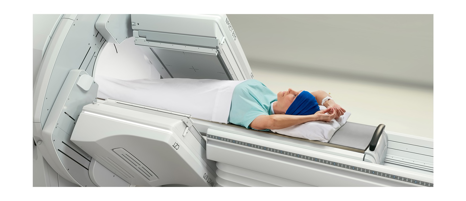
- Patient-friendly scanner designs
Thanks to direct digital conversion and registered collimation, CZT detectors also provide outstanding spatial resolution. With registered collimation, every hole in the collimator is aligned with a single corresponding detector pixel. Registered collimation simplifies localization so events are detected within a single tiny pixel, rather than across a large crystal by multiple PMTs.
More compact, more capable
With no need for PMTs, CZT detectors are far more compact than a NaI-based detector. This compact design paves the way for numerous performance advancements including the ability to position the detector closer to the area to be imaged, further amplifying its exceptional spatial and energy resolutions.
With direct digital conversion and no PMTs in the way, dead space is less of an issue for maintaining close proximity to the patient.
The elimination of PMTs also solves the Anger camera’s inherent design challenge with dead space, or the space at the edge of the detector that isn’t usable because the PMTs overlap the crystal’s edge. With direct digital conversion and no PMTs in the way, dead space is less of an issue for maintaining close proximity to the patient.
CZT evolves to include medium-energy applications
CZT-based detectors capabilities have recently expanded to include the ability to detect the higher energy peaks of medium-energy isotopes. The jump from low- to medium-energy imaging is the result of modifications to two key components of the system.
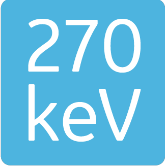
The first of these changes was made to the CZT module itself. Simply put, the optimal imaging of isotopes up to 270 keV requires a thicker CZT crystal. The second set of modifications focused on the collimator. To take advantage of CZT’s enhanced sensitivity over NaI-based imaging at medium energies, changes were made to the collimator’s bore size, shape, length, and material composition.
With these newfound medium-energy imaging capabilities at their disposal, hospitals and imaging centers are now able to apply the image quality, imaging efficiency, and productivity benefits of CZT to their theranostic imaging needs as well.
With these newfound medium-energy imaging capabilities at their disposal, hospitals and imaging centers are now able to apply the image quality, imaging efficiency, and productivity benefits of CZT to their theranostic imaging needs as well.
As medium-energy applications for CZT begin to grow, manufacturers are already conducting research into radiopharmaceuticals and future imaging system designs to help further expand use of tracers that can be imaged with CZT-based systems.
Supporting energies up to 270 keV increases available counts in Theranostic procedures by visualizing both the 113 keV and 208 keV peaks of 177Lu consolidating low- and medium-energy imaging capabilities into a single system will certainly make far more efficient use of your full-service SPECT/CT offering and investment.
Keep exploring to learn more about CZT technology in a premium SPECT/CT camera.


