Regional Medical Imaging Leverages the Power of 3D Mammography and AI to Enhance Breast Cancer Screening
Annual screening mammograms for early breast cancer detection have made a difference in the lives of millions of women. Coupled with the incorporation of artificial intelligence (AI) and advanced imaging, patient outcomes and accuracy have improved[i].
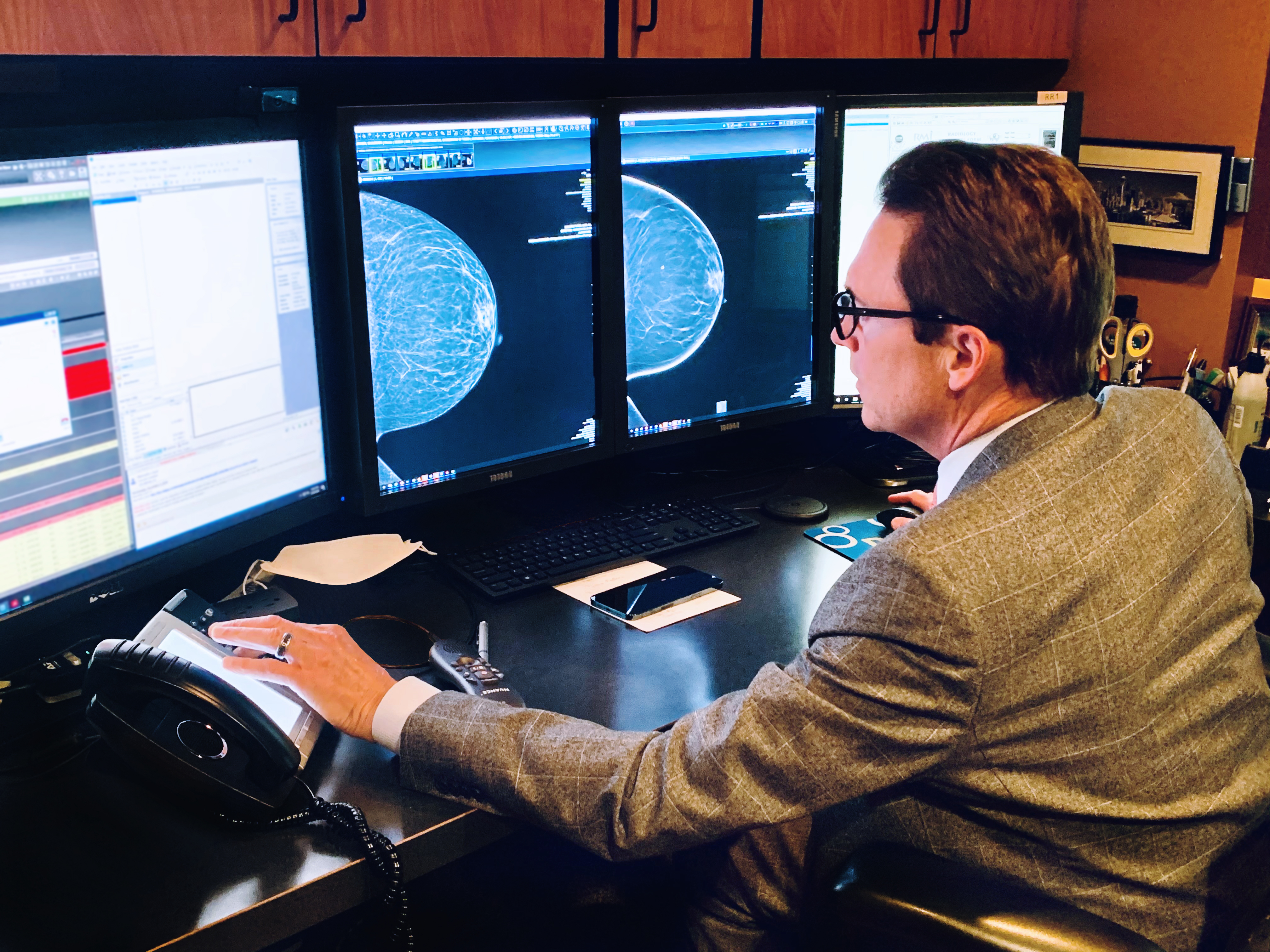
An advocate in the pursuit of excellence in breast health and breast cancer screening, Dr. Randy Hicks, co-owner and CEO of Regional Medical Imaging (RMI) has developed a breast imaging program based on a combination of advanced technology, AI, and experienced clinical staff.
Seeing the impact of 3D mammography
Well-established in the Midwest, RMI has provided imaging services throughout mid- and southern Michigan for more than 35 years. When 3D mammography became available, Dr. Hicks and his team recognized the benefits and felt that it would become a new standard of screening by helping improve breast cancer detection in women with dense breast tissue.
“Adding 3D mammography to our imaging centers really created a buzz around who we are and what we’re doing for our patients,” notes Dr. Hicks. “While 3D is an incrementally small jump in technology, it has allowed us to have the possibility to detect more cancers. Now we participate in several local tumor boards each week and this has enabled us to show our colleagues some of the cancers that would have otherwise been missed. This technology has really elevated our status in our community and certainly has made a difference for women going through this experience.”
RMI elected to install GE Healthcare’s Senographe Pristina™, a mammography platform that can deliver the benefits of 3D technology using the same low radiation doses as traditional 2D mammography[ii]. In addition to its technology enhancements, the Pristina system was specifically designed to create a more relaxing patient experience.
“I love our Pristina,” says Dr. Hicks, “and so do our patients. It’s really a beautiful machine. When patients walk into the room, its warm, soft color, and backlit lighting look inviting to them, and can help ease their anxiety about coming in for their annual screening exam. And in terms of cancer detection, it’s definitely the right direction. 3D mammography is the standard of breast care in most of the U.S. today.”
At the same time RMI made the move to 3D mammography, they also decided to take advantage of some of the powerful AI tools being developed within radiology. The AI reading tools had a profound effect on radiologist workflow, especially with the widespread growth in the adoption of 3D mammography.
“While 3D mammography offers a number of benefits to our patients, such as potentially improved cancer detection with fewer false positives and unnecessary recalls, it yields an exponential increase in the number of images we can read per patient due to the increased visibility, sensitivity, and clinical data it provides,” he said.[iii]
In just three days of reading 3D mammography images, Dr. Hicks said, his radiologists can read the same number of images as in a year of reading 2D images thanks to the ProFound AI technology from ICAD[iv], which acts as a second pair of eyes delivering key data. This, in turn, can help improve radiologists’ confidence in making clinical decisions and prioritizing caseloads, while helping reduce reader fatigue.
Enhancing breast screening with AI
“Using AI, we have absolutely seen excellent results. We know we are helped in finding more cancers, so ultimately, our patients are winning. In one notable example, we saw a young patient with what seemed like a total white-out of the breast. By using 3D imaging and allowing the AI to find the cancer, we were better able to finally spot the cancer when we didn’t see it originally. The powerful combination of these tools gave us the opportunity to identify it. These are exactly the types of cases we’re sharing at the tumor boards,” says Dr. Hicks.
Despite installing sophisticated imaging technology and reading tools, Dr. Hicks believes neither could be effective without the most important diagnostic expertise that can only be found in the reading radiologists.
“You can’t have 3D or AI without the radiologist,” says Dr. Hicks, “but working in synchrony, we make a very strong team.”
In addition, they are able to assist their patients along their care paths with supplemental screening exams such as contrast-enhanced spectral mammography (CESM), automated 3D breast ultrasound (ABUS) and 4D breast MRI, as well as other diagnostic procedures and preoperative imaging.
“In the end, our goal is to find as many cancers as we can as early as we can so they can be treated,” concludes Dr. Hicks. “And so we’re employing the best technology available to help us do that. We’re always trying to improve our skillset for the benefit of our patients.”
[i] Study finds ICAD’s ProFound AI Improves Efficiency and Accuracy in Breast Cancer Detection: https://www.itnonline.com/article/study-finds-icads-profound-ai-improves-efficiency-and-accuracy-breast-cancer-detection
[ii] Data on File. GE Healthcare 2017.
[iii] Superior diagnostic accuracy demonstrated in a reader study comparing the ROC AUC of GE screening protocol (V-Preview + 3D CC/MLO with 3D in STD mode) to that of 2D FFDM alone. V-Preview is the 2D synthesized image generated by GE Seno Iris mammography software from GE DBT images. FDA PMA P130020 https://www.accessdata.fda.gov/scripts/cdrh/cfdocs/cfpma/pma.cfm?id=P130020.
[iv] Compared to reading without ProFound AI. iCAD labelling and user manual, DTM160 rev C. Reading times may vary based on the specific functionality of the viewing application used for interpretation.