Don't have an account?
Critical insights for critical decisions
Learn how GE Healthcare’s cardiology solutions deliver insights that can help you make the decisions that matter for your patients, your clinical staff and your cardiology department.


Patients with Atrial Fibrillation are hospitalized twice as often as patients without AFib and are 3 times more likely to have multiple admissions.1 AFib costs the United States about $26 billion each year.1
Time is critical for patients with Atrial Fibrillation who present with cardiac issues. GE’s Cardiovascular IT and Diagnostic Cardiology solutions can provide insights throughout the cardiology care pathway to help improve clinical decisions and outcomes.


There is a strong link between AF and stroke - one in five of all strokes can be attributed to this condition. Approximately 30 percent of people who experience AF-related strokes die during the hospital admission and another 20 percent are likely to die within a year. Care for patients with Atrial Fibrillation starts with identifying high-risk populations, for example–patients with sleep apnea, obesity and/or hypertension. Some regional guidelines recommend regular screening of all patients over 65 years old.

ECG Recorders
Instant capture medical-grade single-lead EKGs on mobile device.


Patients are typically managed with medical therapy such as anti-coagulation and anti-arrhythmic medications. Cardiovascular imaging can also be employed to help determine cardiac function. For patients who don’t respond to medical therapy alone, there are non-surgical treatment options such as cardioversion and ablation.3
Vivid™ E95
cSound™ beamforming technology for exceptional visualization quality with impressive resolution in 2D, color flow, Doppler and 4D formats.


Intervention
During the intervention, it’s important to minimize complications. Interventional system use should be optimized for clinical success while helping to minimize contrast and radiation exposure to patient and clinician.


Patient Follow-Up
After an ablation, regular patient follow up and non-invasive imaging assessment may detect potential return of Atrial Fibrillation.

Atrial Fibrillation Sources
Time to intervention can be a risk: delays to reperfusion correlate with higher rates of mortality and morbidity. Approximately 80 percent of all CVD deaths are due to heart attacks.1
Time is critical for patients who present with acute chest pain, with or without cardiac history. GE’s Cardiovascular IT and Diagnostic Cardiology solutions can provide insights throughout the cardiology care pathway to help improve clinical decisions and outcomes.
Pre-Hospital Transfer
Rapid transport, ECG monitoring and patient stabilization are critical for patients having an ST-segment elevation myocardial infarction. Nearly half of potentially salvageable myocardium is lost within one hour of the coronary artery being occluded, and nearly 2/3 lost within three hours.3
The ability to send observations and test results wirelessly can be invaluable to attending physicians and interventional lab preparations — potentially enabling fast action upon arrival to aid quality patient outcomes such as door to balloon time.4

Diagnosis
There are many potential causes of chest pain. For patients not sent immediately to the interventional lab for revascularization, guidelines suggest use of biomarkers, clinical history, ECG and non-invasive imaging measurement results, and other factors, to determine TIMI risk score which in turn can guide revascularization decisions.5
There are available estimates for sensitivity and specificity for the various non-invasive tests to diagnose the presence of CAD, but other factors are frequently considered such as equipment availability and patient tolerance.6


Intervention
For patients sent for revascularization in the catheterization lab, attempts are made to rapidly access the culprit artery and complete further assessment of coronary tree. Rapid intervention can help minimize time to reperfusion from first medical contact.


Patient Follow-Up
Follow-up treatment and rehabilitation strategies typically include non-invasive monitoring of LV function and strain.


Acute Coronary Syndrome Sources:
Among symptomatic patients with medically treated moderate-to-severe aortic stenosis, mortality from the onset of symptoms is approximately 25 percent at one year and 50 percent at two years.1 GE Healthcare Cardiology Solutions help you make decisions that matter for your patients, staff and hospital.
Time is critical for AS patients who present with significant cardiac issues. GE’s Cardiovascular IT and Diagnostic Cardiology solutions can provide insights throughout the cardiology care pathway to help improve clinical decisions and outcomes.
Diagnosis involves assessment of valve morphology, amount of stenosis or insufficiency, LV function, LV hypertrophy, and severity of symptoms. For patients with Aortic Stenosis, there are surgical and catheterization treatment options in addition to medical or palliative care. A risk calculator is typically used to quantify the risk and make a treatment decision.


For patients choosing a transcatheter replacement, non-invasive cardiovascular imaging, such as Ultrasound, CT, and MR are typically used to assess anatomy to plan the access site and size the valve.


During the transcatheter aortic valve replacement, steps are taken to ensure the most effective result, including valve sizing, position, and function are optimal.


After the procedure, care is taken to ensure functionality of the valve typically using non-invasive imaging and ECG.

Aortic Stenosis Sources:
Managing risk factors and follow up for patients with Stable Ischemic Heart Disease is critical. GE’s Cardiovascular IT and Diagnostic Cardiology solutions can provide insights throughout the cardiology care pathway to help improve clinical decisions and outcomes.
Early detection and management including counseling, and medical therapy are important for patients with cardiovascular disease or who are at a high risk, including those with risk factors such as hypertension, diabetes, and high cholesterol.1
Non-invasive methods for observing response to guideline-directed therapy can be employed, two examples of these methods are 12 SL ECG interpretation and stress imaging. Additionally, if the patient is not responsive to therapy, CT, MR or Nuclear studies may help show functional degradation which might necessitate a follow on intervention such as PCI or CABG.


Successful PCI of the stenotic lesions involves planning, guidance, and assessment of the culprit lesion and assessment of the remainder of the coronary tree.


Follow-up treatment and rehabilitation strategy can include behavior modification and medical therapy in addition to monitoring cardiac function non-invasively using ALARA principles.

Stable Ischemic Heart Disease Sources:
Heart Failure cost is 2-3 percent of the total health-care expenditures in high-income countries, and costs are projected to increase 200 percent in next 20 years world-wide. Current cost in the U.S. is $30.7 billion dollars.1 Congestive heart failure affects people of all ages, from children and young adults to the middle-aged and the elderly.
Time is critical for Heart Failure patients who present with cardiac issues. GE’s Cardiovascular IT and Diagnostic Cardiology solutions can provide insights throughout the cardiology care pathway to help improve clinical decisions and outcomes.
About half of people who develop heart failure die within five years of diagnosis.3 Important risk factors include HTN, DM, Metabolic Syndrome, and Coronary Artery Disease, but there are many other conditions that put patients at risk. Early identification and treatment of comorbidities can delay the onset of Heart Failure.4

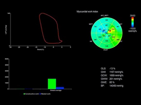
Diagnosis typically involves assessment for ECG abnormality and assessment of cardiac structure, function and possible cause. Guidelines suggest use of echocardiography due to its widespread availability, but other non-invasive imaging modalities can be used. In certain cases, invasive monitoring provides useful information for patients with persistent symptoms. Biomarkers provide helpful information about progression of disease. 4


Guidelines suggest lifestyle modifications, and use of specific medical therapies, such as ACE inhibitors and beta blockers. For certain patients not responding to medical therapies, guidelines suggest use of ICD or CRT. Revascularization and valve repair are used in certain stages of HF.4
Guidelines recommend regular follow up to assess severity and extent of left ventricular dysfunction, and development of a follow-up treatment strategy, including monitoring, to prevent acute decompensation and re-admission.4


Heart Failure Sources:
Flexible offerings can help ensure optimal equipment and clinical performance, and includes tools and resources to optimize uptime to reduce patient disruptions. With the ability to add services beyond maintenance, clinical confidence is further supported through remote and on-site applications training and support.

We help clients solve the most challenging problems in healthcare through long-term strategic partnerships, and advanced analytic capabilities. We collaborate to define and prioritize their critical challenges, design the best strategies, and activate impactful solutions to create breakthrough, sustainable outcomes to enable them to transform and succeed.
