Don't have an account?
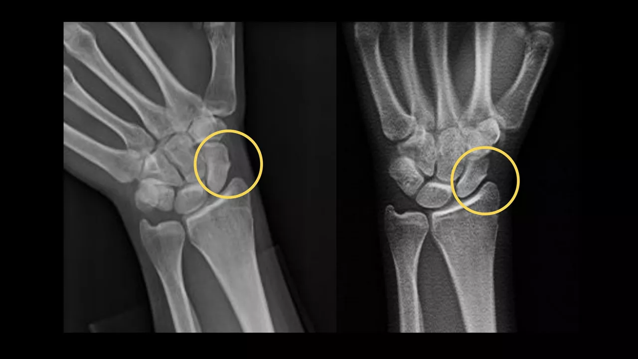
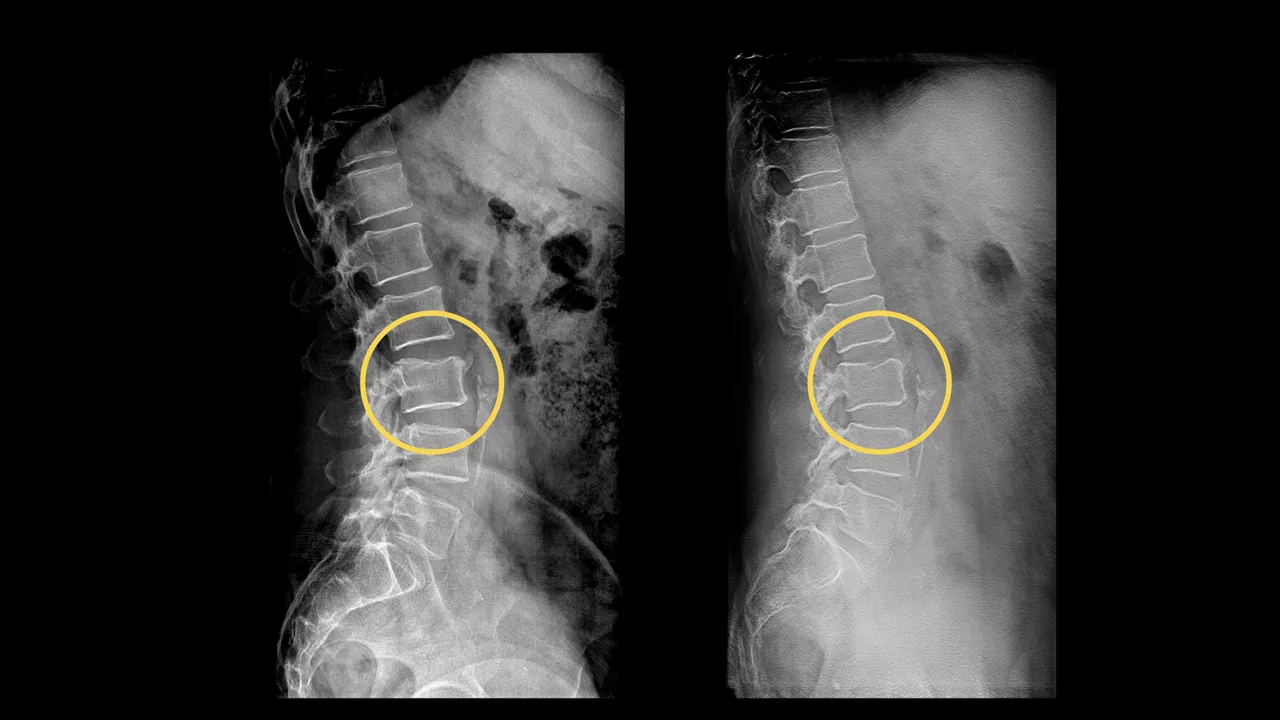
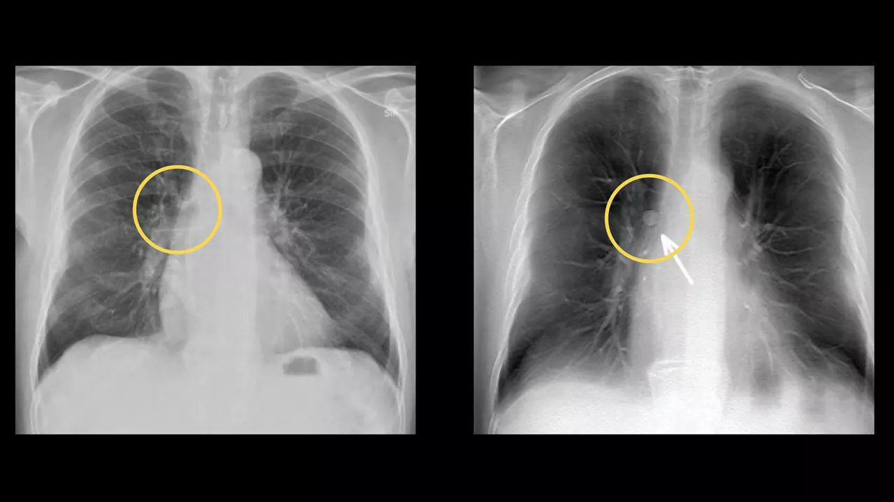
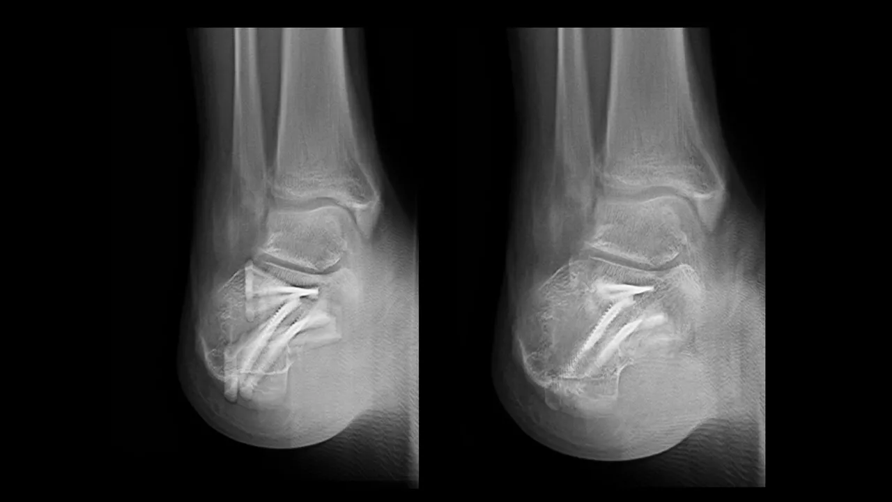
When an X-ray is inconclusive, VolumeRAD™ provides additional information to assist radiologists with confident, diagnostic decision-making. The exam can be performed in <2 minutes at the same time and same system—with 20x less dose than a CT exam.1-5