• Change country/language

    Looks like you’re in {} — you’re on the {} site.

    Cancel

    Country and language selected

    You are being redirected to the {} site. Products and services availability vary by country. Do you wish to continue?

    Continue
    Cancel
Nuclear Medicine

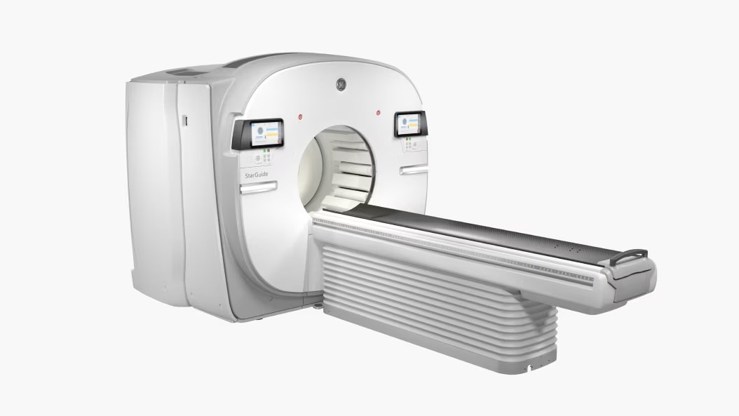
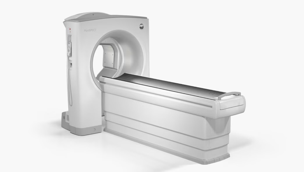
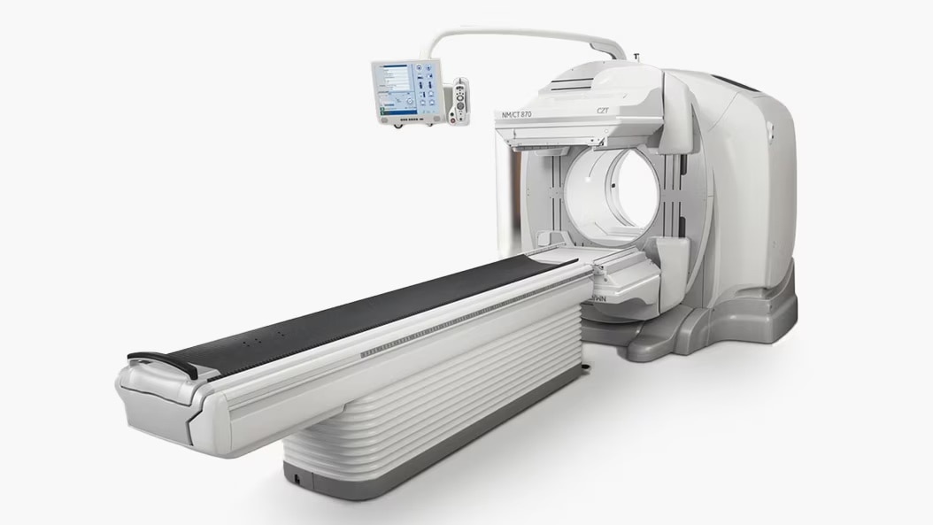
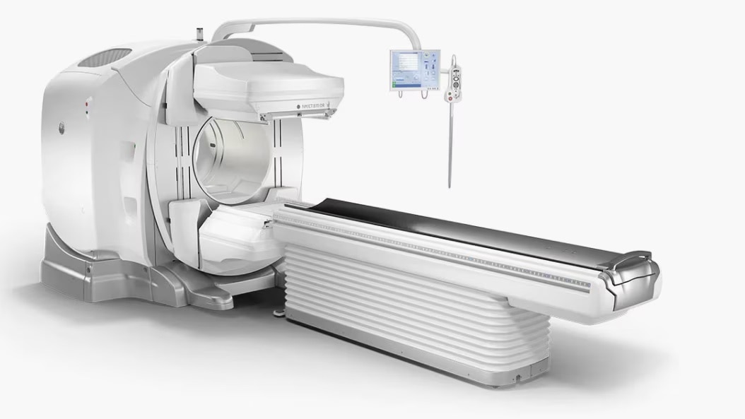
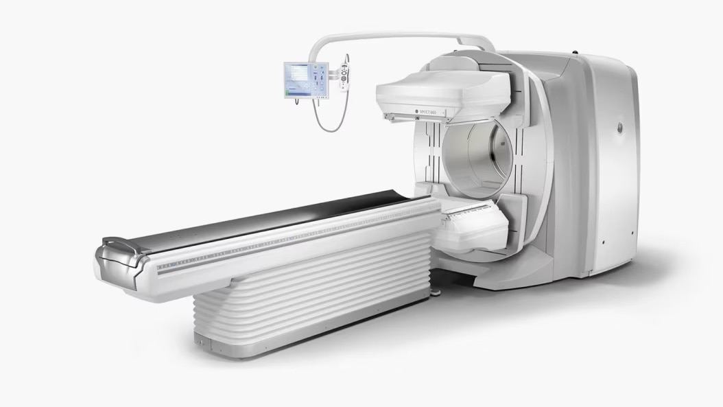
With a wide range of available isotopes and the flexibility to explore multiple pathways in a single session, NM cameras are full of potential. Our latest advancements are making nuclear medicine technology more accessible and enabling it to provide you with more meaningful clinical answers.

StarGuide™

Introducing StarGuide, our most advanced SPECT/CT featuring advanced Cadmium Zinc Telluride (CZT) technology.

Explore

New protocols can be visualized with multiple tracers simultaneously.

Search

New insights with 3D Dynamic Acquisition¹ can be searched.

Research

Existing tracers help expand clinical applications.

Support

Assisting efforts for the development of new tracers.

MyoSPECT™

MyoSPECT Molecular Imaging was designed with the same focused attention to the heart that defines the entire practice of cardiology.

Clear, more complete look at every patient's heart

Extended FOV processing for a 76% increase in FOV volume²

True dynamic imaging with SPECT Flow

Combine dynamic acquisition on MyoSPECT with CFR and aboslute myocardial blood flow

Two different attenuation compensation solutions

Evaluate attenuation by imaging in prone and supine positions, or combine the perfusion images with separate CT images on Xeleris

Smart Positioning Workflow

Automated scan position and FOV recommendations

NM/CT 870 CZT

NM/CT 870 CZT is the third-generation of our general purpose digital SPECT/CT system powered by CZT technology.

Improved lesion detectability³

Assisting efforts to diagnose, stage disease earlier

Guide therapy planning decisions

With quantitative disease state and treatment response assessments

Theranostics capabilities

Use to enhance your full-service SPECT/CT practice

Productivity

Simplified workflows for complex procedures

NM/CT 870 DR

NM/CT 870 DR, a leading light in clinical discovery

Uncompromised image quality

Improved small lesion detectability with SwiftScan Planar and SwiftScan SPECT4

Ready for Theranostics

Only scanner with easy, direct upgrade to CZT technology

Enhance productivity

Simplify workflows for complex procedures with SmartConsole

AI solutions

Several quantitation-based applications, powered by AI on Xeleris

NM/CT 860

NM/CT 860 is a SPECT/CT system designed for high-performance clinical environments combining the SPECT and 800 Series with an optimal balance of CT technology.

Save time and steps

by remotely collaborating with a clinician mid-exam

Enhance productivity

with simplified workflows for complex procedures

Easy-to-use user interface

helps your department operate efficiently

Improve workflow

with automated transfer and archive features

NM/CT 850

NM/CT 850 is our most accessible SPECT/CT system. It combines the SPECT image quality and productivity enhancements of the 800 Series with the essential CT technology for providing that all-important layer of anatomical information, which you need specifically for localization and attenuation correction in SPECT imaging.

Our most accessible SPECT/CT system

Has the smallest footprint of all our 800 Series SPECT/CT systems

Enhance productivity

with simplified workflows for complex procedures

Increase confidence in your images

Leveraging the CT technology you need for anatomical localization and attenuation correction

Cost-effective system upgrade

Shares the same modular design as the rest of the 800 Series family of nuclear medicine systems

NM 830

NM 830 SPECT/CT Sytem brings you the diagnostic capability of a dual-head SPECT system with a focus on patient comfort.

Enable reduction of dose or scan times

by up to 25 percent with the increased sensitivity of SwiftScan Planar and SwiftScan SPECT5

Shorter, more tolerable exams

for greater patient comfort, with Evolution technology6

Diagnose disease earlier

with SwiftScan Planar and SwiftScan SPECT’s improved small lesion detectability4

CZT: Precision imaging at the speed of tomorrow

CZT is the key component to the future of nuclear medicine. Not only is it lighter and more compact than the Nal crystals used in analog technology, it is pixelated. With registered collimation, each photon is directly converted into an electrical signal that more accurately identifies its location and energy. The result is increased spatial and energy resolution.

nuclear-medicine-czt-ph-en


Disclaimers
  1. Acquisition only. The acquired dynamic data is stored in a list file.
  2. As compared to Discovery™ NM 530c.
  3. In clinical practice, the use of NM/CT CZT 870 with WEHR collimator may improve lesion detectability depending on the clinical task, patient size, anatomical location and clinical practice. A consultation with a radiologist and a physicist should be made to determine the appropriate dose or scan time to obtain diagnostic image quality for the particular clinical task.
  4. As demonstrated in phantom testing using a model observer. For SPECT, compared to using the LEHR Collimator and a SPECT Step & Shoot acquisition. For Planar, compared to using LEHR without Clarity 2D.
  5. Compared to LEHR collimator, with Step & Shoot scan mode (for SPECT) / without Clarity 2D (for Planar). As demonstrated in phantom testing using a bone scan protocol, Evolution processing (for SPECT), and a model observer. Because model observer results may not always match those from a human reader, the actual time/dose reduction depends on the clinical task, patient size, anatomical location and clinical practice. A radiologist should determine the appropriate scan time/dose for the particular task.
  6. In clinical practice, Evolution options6a (Evolution for Bone, Evolution for Cardiac, Evolution for Bone Planar) and Evolution Toolkit6b are recommended for use following consultation of a Nuclear Medicine physician, physicist and/or application specialist to determine the appropriate dose or scan time reduction to obtain diagnostic image quality for a particular clinical task, depending on the protocol adopted by the clinical site.

    6a. Evolution Options - Evolution claims are supported by simulation of count statistics using default factory protocols and imaging of 99mTc based radiotracers with LEHR collimator on anthropomorphic phantom or realistic NCAT - SIMSET phantom followed by quantitative and qualitative images comparison.
    6b. Evolution Toolkit - Evolution Toolkit claims are supported by simulation of full count statistics using lesion simulation phantom images based on various radiotracers and collimators and by showing that SPECT image quality reconstructed with Evolution Toolkit provide equivalent clinical information but have better signal-to-noise, contrast, and lesion resolution compared to the images reconstructed with FBP / OSEM.


JB23688XX May 2023

Have a question? We would love to hear from you.

Contact us

We're ready to support you in your moments that matter.

We’re sorry, a problem occurred with the form. Please try again.
Thank you for your inquiry.

We will be in touch soon.

Something went wrong

We will check and get back.