Don't have an account?
Access to spectral CT information without impacting your clinical workflow.
At a glance
Seamless integration
Access to spectral CT information without impacting your clinical workflow
Advanced visualization
Leverage the power of spectral imaging with 3D advanced visualization capabilities
Dedicated protocols
Automate reviews: Gout protocol, Pulmonary Perfusion1 or Liver Fat2
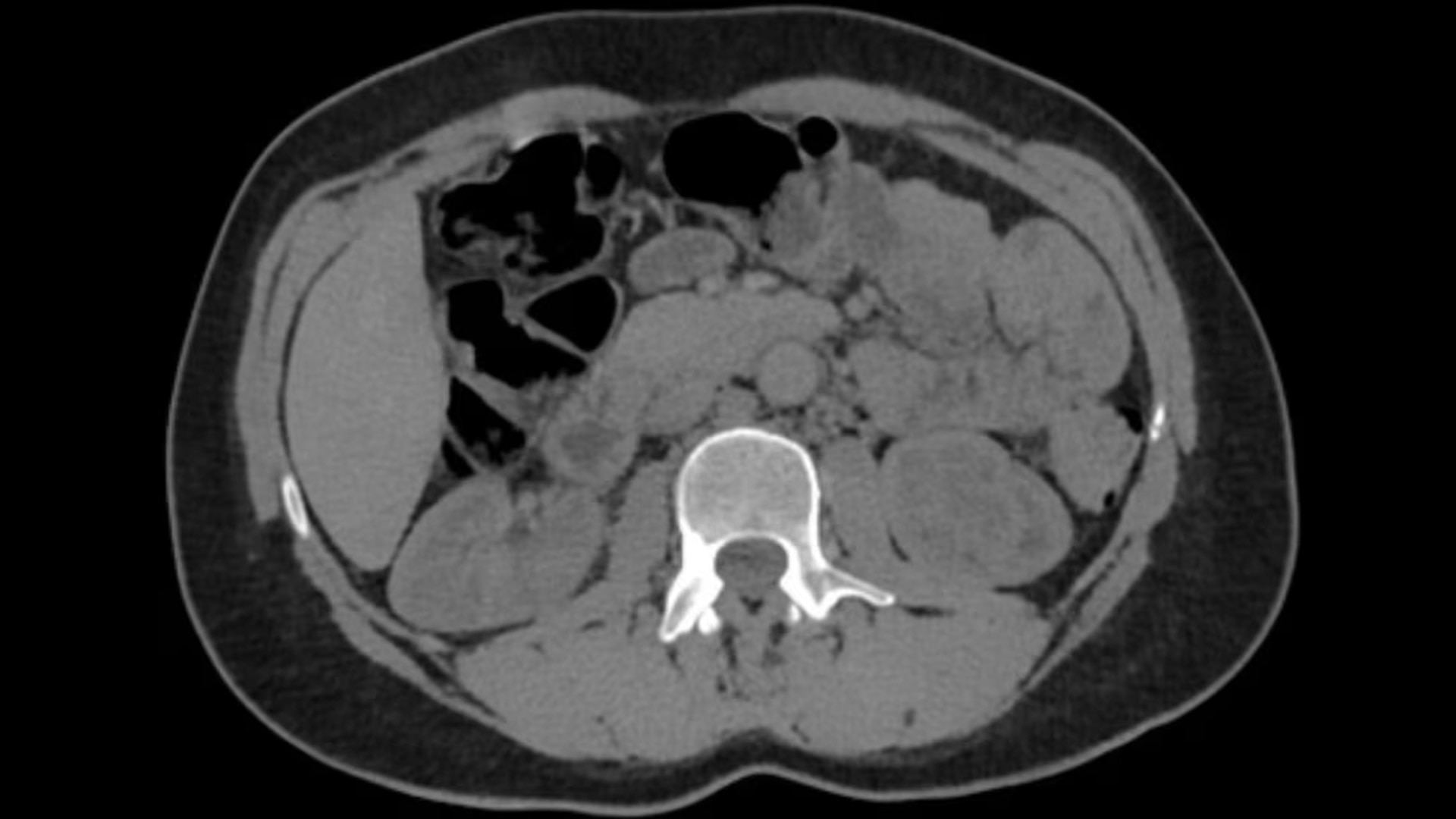
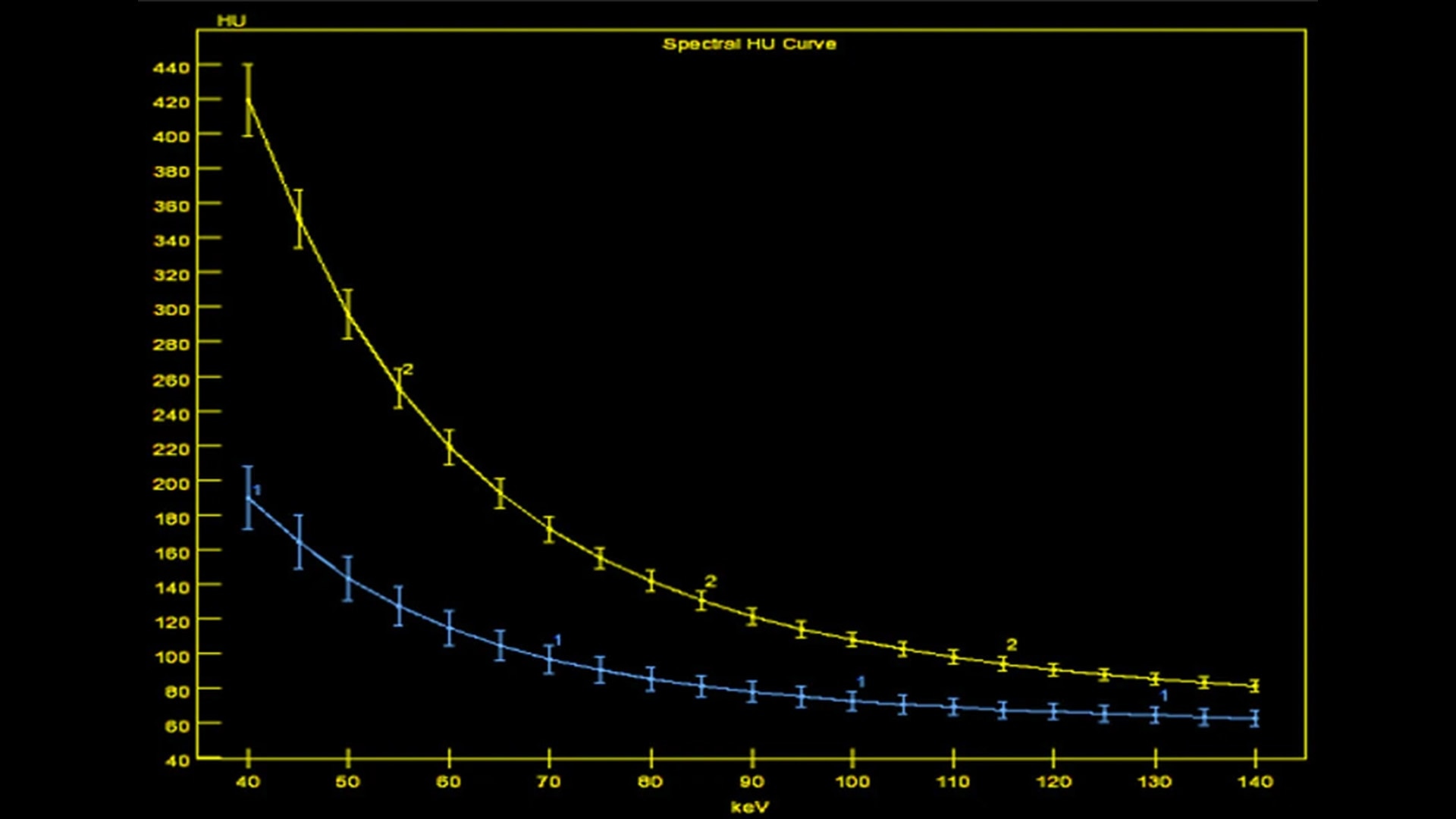
GSI Viewer allows for the review of monochromatic energy images at user selectable energy levels, detailed analysis using material decomposed images, and complementary information using the Effective-Z images.
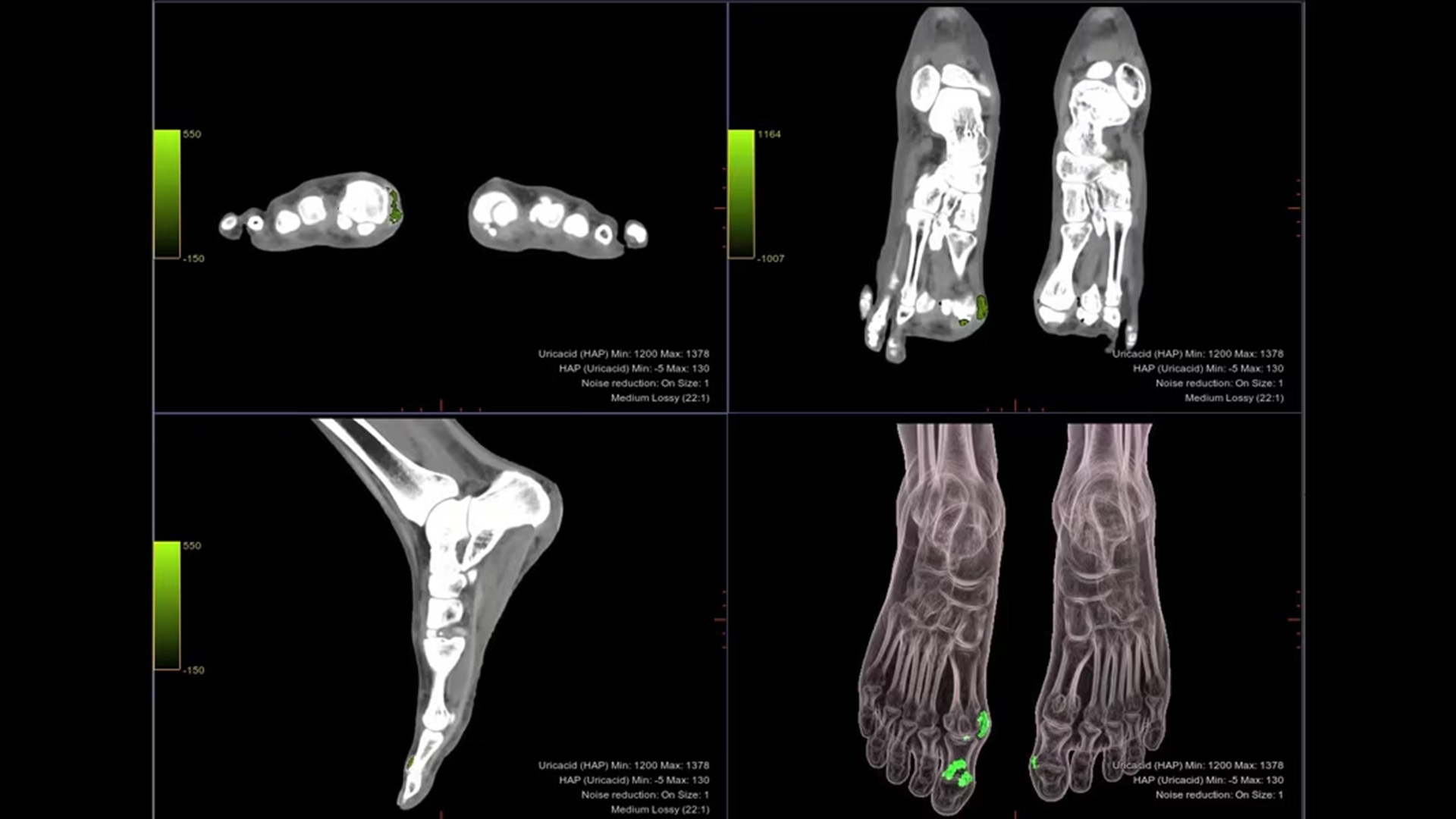
Fully automated workflow for material decomposition and color overlay to assist in gout characterization:
Automated generation of 2D and 3D gout overlay
Automated export of 2D and 3D gout overlay images to the patient browser
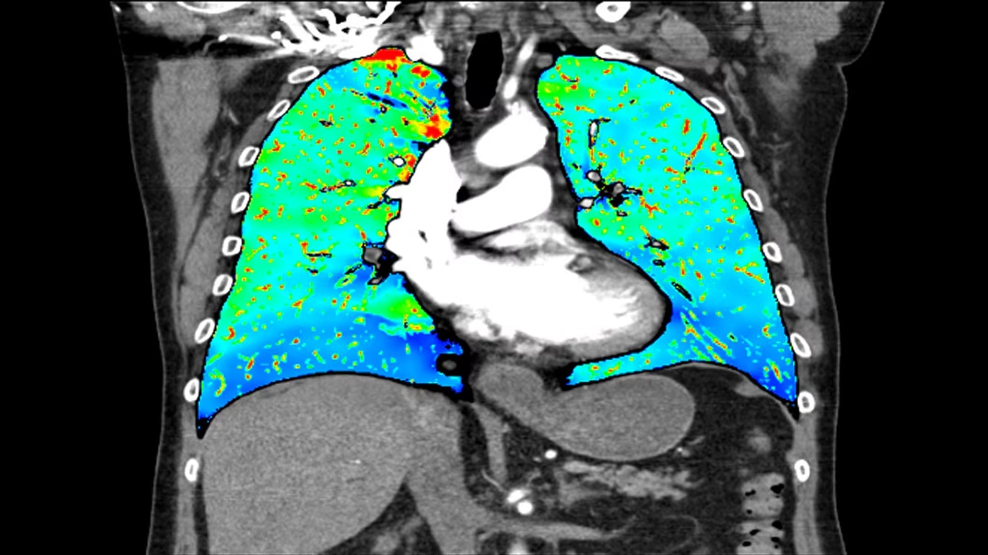
One-click protocol for relative iodine distribution
Automatic relative hypo-perfused region display
Volumes reported for total lung and relative hypo-perfusion volumes
Succinctly communicate volumes and relevant pictures to referring physicians
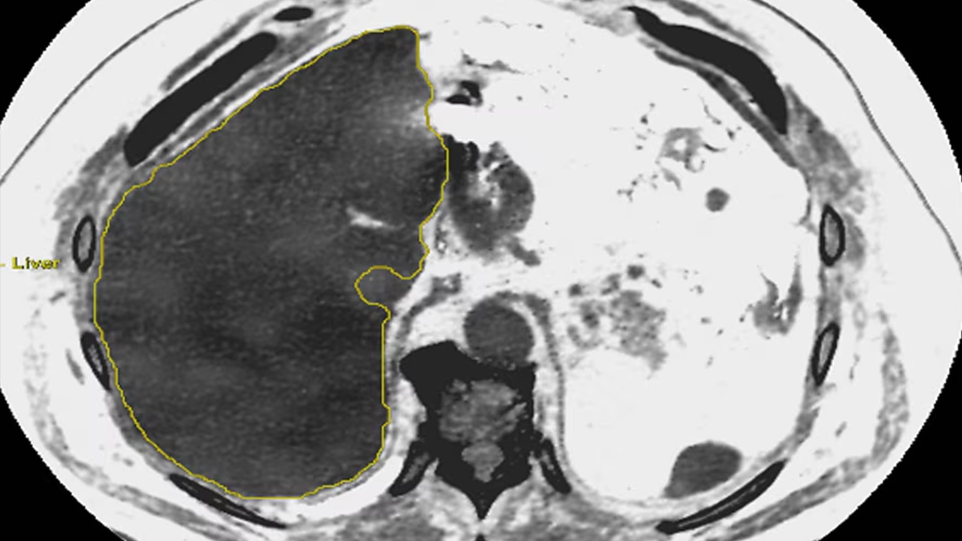
Dedicated GSI Fat capability to provide approximate percentage of fat in the liver:
Help evaluate hepatic steatosis with liver fat percentage measurements
GSI with material separation provides the ability to measure approximate liver fat percentages
Liver fat measurements compatible with non-contrast and multi-phase contrast acquisitions