• Change country/language

    Looks like you’re in {} — you’re on the {} site.

    Cancel

    Country and language selected

    You are being redirected to the {} site. Products and services availability vary by country. Do you wish to continue?

    Continue
    Cancel
VersaViewer™ with One View

AI-optimized spectral CT visualization

At a glance

Reduce complexity

Access all relevant information in one clear summary view — no manual window adjustments, no switching between spectral datasets.

Automate precision

See each organ with automatically optimized windowing for fast, intuitive interpretation.

Streamline Spectral CT

Unlock the benefits of spectral data with OneView, accessible from the web client.


Transforming spectral complexity into clinical clarity

Radiologists face challenges with complex multi-energy datasets and cumbersome workflows of Spectral CT. One View unifies Spectral CT datasets into a single, AI-optimized view to help support faster, more accurate diagnoses.
Key features
Advanced AI features for spectral CT interpretation and workflow efficiency
Multi-organ segmentation with AI
See more. Automatically.
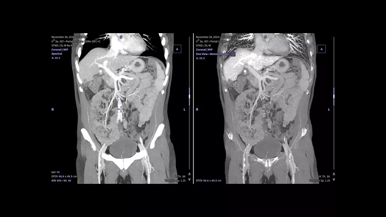
AI-based algorithms segment multiple organs1 and anatomical regions in the torso, including lung, liver, bone, aorta and heart. By automatically identifying structures, clinicians can focus more on image interpretation rather than manual delineation.
Clinician reviewing a coronal abdominal CT scan with color-coded AI segmentation on a workstation monitor.
Automatic contrast optimization
Optimized for every organ
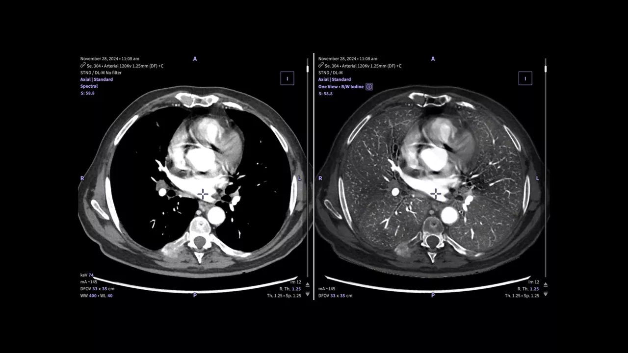
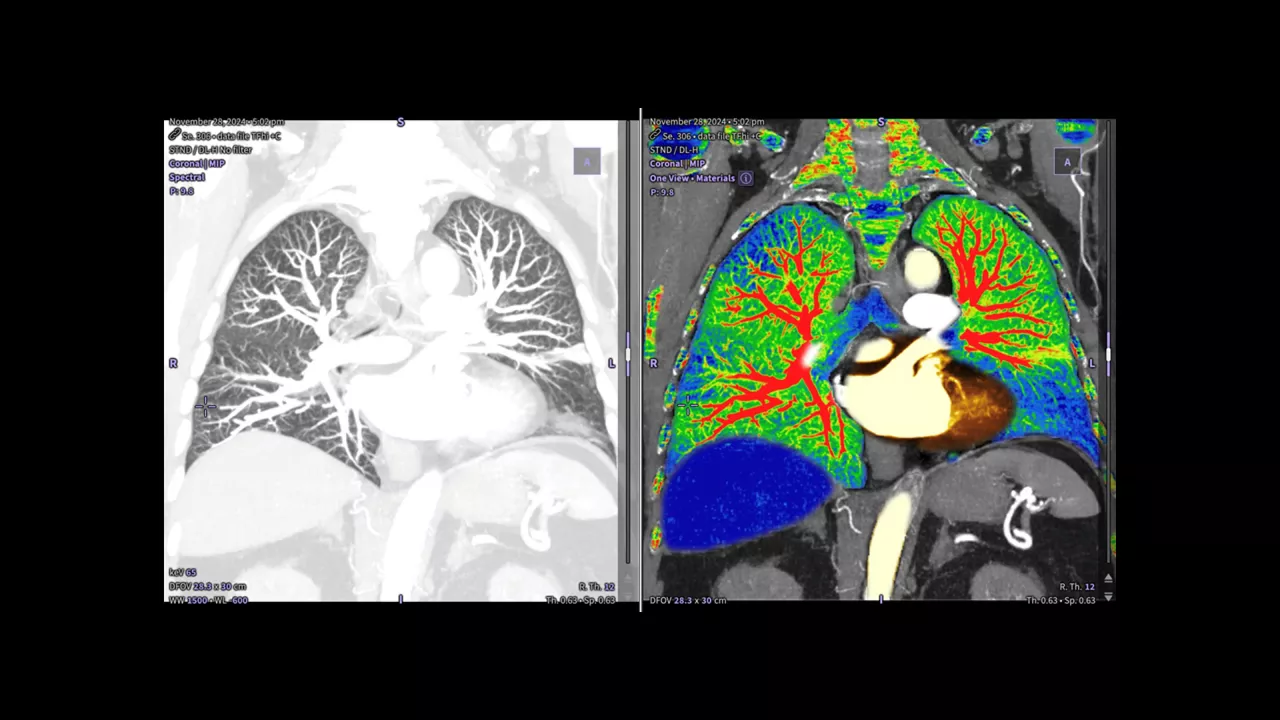
Contrast parameters are automatically adjusted by organ to support visualization and diagnostic review. Presets are available in both color and grayscale to enable intuitive interpretation with fewer manual adjustments.
Color-coded axial chest CT showing optimized contrast across the lungs, heart and surrounding anatomy.
Unified spectral visualization
Consolidated spectral insights in one intelligent view
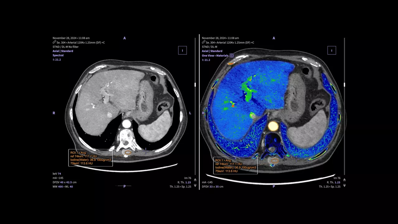
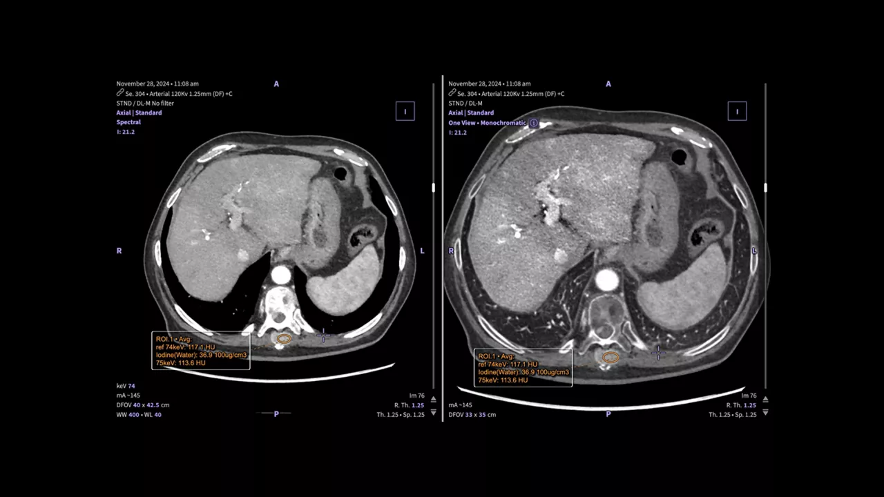
One View consolidates spectral information2 into a single, AI-optimized image while displaying it alongside the original volume for concurrent review. This unified presentation eliminates the need to switch between datasets, supporting clinicians with comprehensive insight with greater efficiency.
Side-by-side axial abdominal CT scans comparing the original grayscale volume with a color-coded spectral view of the liver.
Disclaimers
  1. Lung, liver, bone, aorta, heart and body (entire body)

  2. Optimized MD and keV by organ
JB39632XX October 2026
Get in touch

Have a question? We would love to hear from you.

Contact us

We're ready to support you in your moments that matter.

We're sorry, a problem occurred with the form. Please try again.
Thank you for your inquiry

We will be in touch soon.

Something went wrong

We will check and get back.