• Change country/language

    Looks like you’re in {} — you’re on the {} site.

    Cancel

    Country and language selected

    You are being redirected to the {} site. Products and services availability vary by country. Do you wish to continue?

    Continue
    Cancel

FastStroke provides a comprehensive workflow solution for reviewing stroke workup images with exceptional flexibility, simplicity and performance. It is a streamlined approach that smartly adapts to your scan practices and allows you to review and post-process all your images simultaneously.


The application provides quick loading and clinically relevant organization of all the scanned series, which are synchronized and displayed in a manner that enables you to review, efficiently and with high confidence.


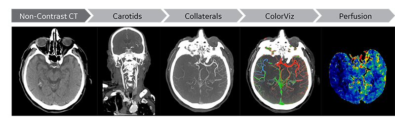
FastStroke also provides ColorViz to aid in the visualization of the timing of collateral vessels using the mCTA series. FastStroke has full integration with CT Perfusion 4D to provide automatic neuro perfusion analysis as part of the workflow.

Features

Features

  • Easy and intuitive workflow to quickly access all series of a CT stroke workup simultaneously
  • Flexible layouts that automatically adapt to display all acquired phases within the mCTA
  • Intelligent color coded display provided by ColorViz. this enables an easy and confident identification of vascular enhancement timing, including collaterals, in a single composite view.
  • Fully integrated single workflow with Neuro Perfusion automated processing

System Requirements

  • AW VolumeShare 7 or later
  • AW Server 3.2 or later
  • Color Monitors
  • Single or Dual Display Monitors for AW Server in Landscape and Portrait orientations

Intended Use

FastStroke is a CT image analysis software package that assists in the analysis and visualization of CT data derived from DICOM 3.0 compliant CT scans. FastStroke is intended for the purpose of displaying vasculature of the head and neck at different time points of enhancement. The software will assist the user by providing optimized display settings to enable fast review of the images in synchronized formats, aligning the display of the images to the order of the scans and linking together multiple groups of scans.


In addition, the software fuses the vascular information from different time points into a single colorized view. This multiphase information can aid the physician in visualizing the presence or absence of collateral vessels in the brain. Collateral vessel information may aid the physician in the evaluation of stroke patients.

Find More Imaging Software by Modality

CT Advanced Visualization
ico-laptop
Interventional Advanced Visualization
MR Advanced Visualization
PET Advanced Visualization

Find More Imaging Software by Clinical Area

Cardiology Advanced Visualization
Oncology Advanced Visualization
RT Advanced Visualization
ico-settings
Vascular Advanced Visualization
Contact Us

We're ready to support you in your moments that matter. For service assistance, call us.

Thank you!

We received your request for assistance.

A GE Healthcare specialist will be reaching out shortly.

Something went wrong

We will check and get back.

JB01331IN March 2023