• Change country/language

    Looks like you’re in {} — you’re on the {} site.

    Cancel

    Country and language selected

    You are being redirected to the {} site. Products and services availability vary by country. Do you wish to continue?

    Continue
    Cancel
BrainWave

At a glance

Non-invasive

No ingestion or infusion of tagged material required, and ionizing radiation free

Generated maps

Yields information that can help guide surgical intervention procedure

Easy-to-use analysis

Enables processing, analysis, 3D rendering and display of results

Visualization tool

Measures heightened oxygen levels in response to external stimuli

Highlights

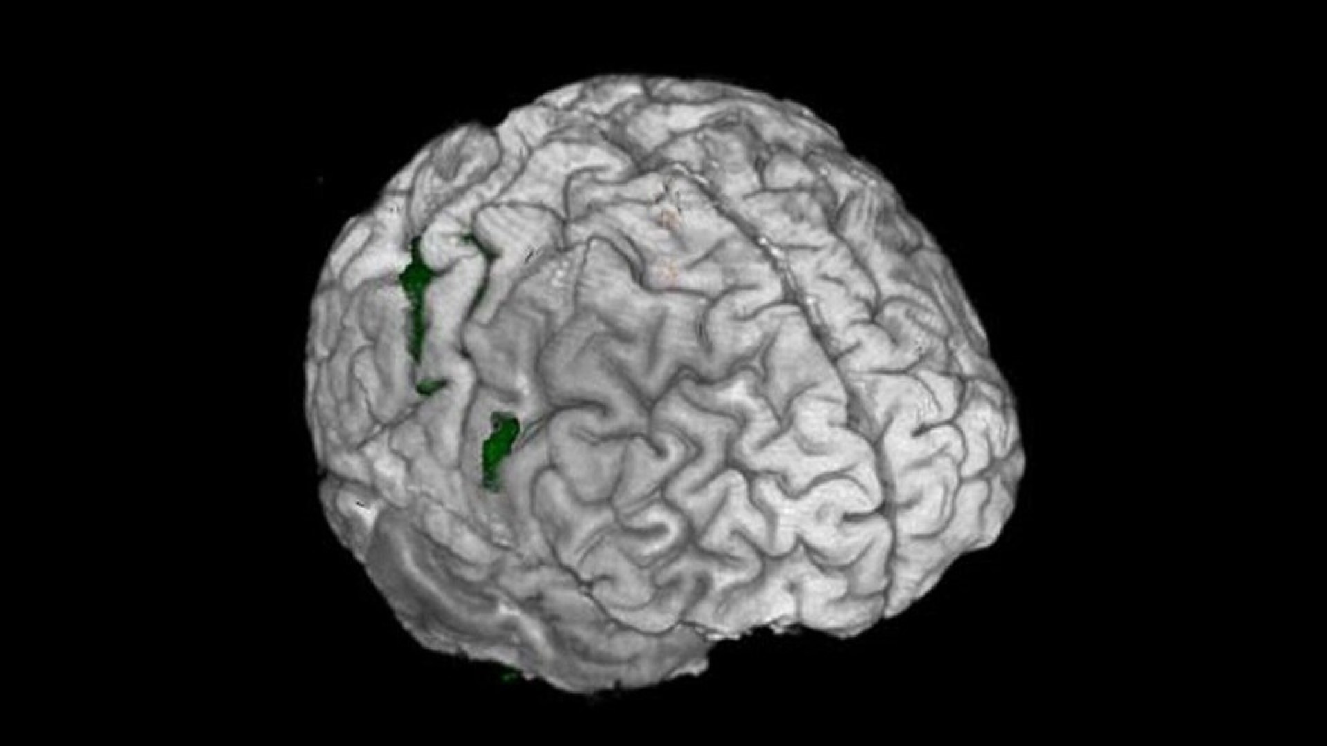
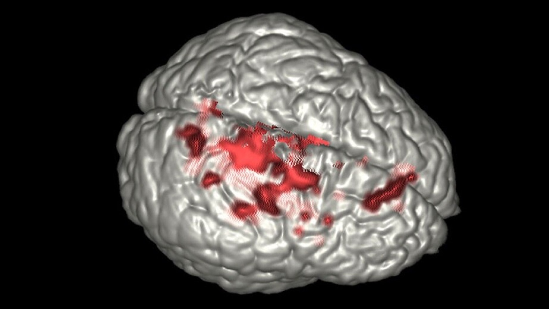
BrainWave imaging software is an easy-to-use analysis and visualization tool for functional brain image data acquired with BrainWave RT. BrainWave enables processing, analysis, 3D rendering and display of results from BOLD MRI scans. It provides:


  • Retrospective motion correction
  • Activation maps generated using Generalized Lineal Model (GLM) analysis
  • Sophisticated visualization techniques that fuse analysis results with anatomical data
  • Clear visualization with color activation maps and interactive thresholding
  • Paradigms saved in the DICOM private structure for fast analysis
  • Full integration with AW filming, networking, and archiving
  • Full DICOM fidelity
  • Ability to transfer activations into high resolution 3D DICOM data sets for neurological applications
Features
  • BrainWave PA lets you interactively view and edit fMRI data. It can reside on the scanner or on your AW station.
  • Software reads volumetric T1-weighted acquisitions in addition to fMRI acquisitions.
  • Processing steps for fMRI data include:

    • Six parameter rigid body motion correction via registration to the first volume.
    • Spatial filtering.
    • Segmentation of the anatomical data so that outer layers of non-brain signal are removed.
    • Registration of fMRI data sets to the structural data set.
    • Calculation of statistical maps corresponding to task-based activation.
    • Formation of Burnt In Pixel (BIP) maps outlining the area of activation in white on the structural scan.
    • Calculation of DTI fiber tracks (option with BrainWave Fusion).
  • View functional data overlaid on the structural image in 2D or 3D renderings. Add annotations or change color and opacities.
  • BrainWave Fusion (option) lets you display white matter tracks with functional data on a high-resolution anatomical image.


features-brainwave-imaging-ci-en
Get in touch

Have a question? We would love to hear from you.

Contact us

We're ready to support you in your moments that matter.

We’re sorry, a problem occurred with the form. Please try again.
Thank you for your inquiry.

We will be in touch soon.

Something went wrong

We will check and get back.

JB29252XX June 2024