A partner you can trust
A complete solution
An opportunity for growth
B-flow / B-flow Color
3D/4D
TVI
Virtual Convex
Elastography for tissue differentiation
Bi-plane imaging
Power Doppler Imaging
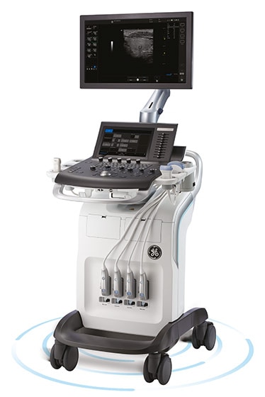
Versana Premier at a glance
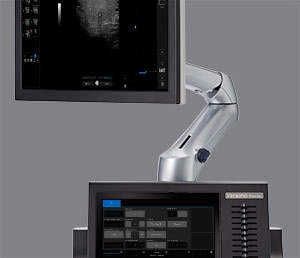
Articulating monitor arm
Integrated battery
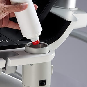
Gel Warmer
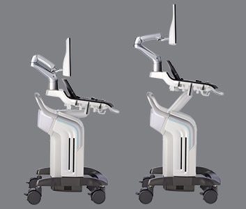
Height-adjustable console
Large full HD display
Touch Panel
Fast facts
- Whizz image tuning
Just touch the Whizz button on your console once. Without pressing it again, Whizz continuously and automatically optimizes the image, even as you move from one organ or structure to another. - Scan Assistant
Provide ultrasound sonographers with predefined standardized exam protocols that walk them through all steps of image acquisition, saving keystrokes, reducing stress and fatigue, and maximizing exam consistency. - Whizz bladder
Use the AutoBladder function within the Whizz feature to combine optimized ultrasound image quality with automated measurements. - Breast Care
Scan breasts with a systematic approach to ensure coverage of all segments; make assessments step by step. For serial exams, compare images to reassess quadrants previously flagged as risks. - Auto EF
Automatically track myocardial tissue deformation and calculate the left ventricular ejection fraction. The system makes the tracings from the apical four-chamber and apical two-chamber views and calculates the ejection fraction. - Needle Recognition
Perform accurate biopsies with technology that clarifies the precise location of the needle point. - Voice comments
Connect a microphone, touch a screen icon, and capture hand-free recorded voice comments that are overlaid on images and saved for playback later when reviewing the exam. - Breast Productivity
Assess breasts while scanning; describe structure characteristics including Bi-Rads in comments that feed directly into the report. Use the Auto Contour feature to easily measure structures. - SonoBiometry
Receive suggested caliper placements to perform standardized BPD, HC, AC, FL and HL fetal measurements. - Insite Technology
GE advanced remote service technology and remote monitoring diagnostics and repair; instantly connect your ultrasound machine with a GE service expert to resolve many issues remotely on the spot. - Stress echo
A template editor for myocardium stress testing. Evaluate heart segment function through myocardium stress scoring and comparison. Stress echo support enables wall motion scoring and automatic stress level labeling of measurements. - Thyroid productivity
Assess the thyroid while scanning; describe structure characteristics in comments that feed directly into the report. - VOCAL
. 3D tool that enables volume calculation of eccentrically shaped areas or complex anatomical structures.
.
.
My Trainer
Scan Coach
Supporting Materials Listing
Products mentioned in the material may be subject to government regulation and may not be available in all countries. Shipment and the effective sale in certain countries can only occur if the product is approved.
Final product configuration and features may differ from the ones represented in the photo/video and may not be available in every region. Check with your local GE representative for details.
© 2020 General Electric Company – All rights reserved. GE Healthcare reserves the right to make changes in specifications and features shown herein, or discontinue the product described at any time without notice or obligation. Contact your GE Healthcare representative for the most current information. GE, GE Monogram, Versana Premier, InSite, CrossXBeam and B-Flow are trademarks of General Electric Company.
GE Healthcare, a division of General Electric Company. GE Medical Systems, Inc., doing business as GE Healthcare.