• Change country/language

    Looks like you’re in {} — you’re on the {} site.

    Cancel

    Country and language selected

    You are being redirected to the {} site. Products and services availability vary by country. Do you wish to continue?

    Continue
    Cancel

Benefits


Discover how University Hospital Donostia and
Jena University Hospital improved their ED practices

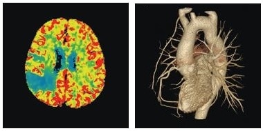
Jena University hospital in Germany upgraded their emergency department (ED) with a Revolution™ CT and ASiR-V™1, GE Healthcare's blended Iterative Reconstruction algorithm. Empowered by these technologies, the clinic has created a new set of clinical protocols called "the Big Five" that combine stroke and chest pain management in the ED to further enhance patient care and increase clinical confidence.


Unicersity Hospital Donostia in Spain installed Revolution™ EVO and ASiR-V to increase efficiency and accuracy in CT scans while lowering dose for thousands of stroke and polytrauma patients each year.



Disclaimer


1. In clinical practice, the use of ASiR, ASiR-V and Veo may reduce CT patient dose depending on the clinical task, patient size, anatomical location, and clinical practice. A consultation with a radiologist and a physician should be made to determine the appropriate dose to obtain diagnostic image quality for the particular clinical task. ASiR-V low contrast detectability (LCD), image noise, spatial resolution, and artifact were assessed using reference factory protocols comparing ASiR-V and FBP.

The LCD measured in 0.625 mm slices and tested for both head and body modes using the MITA CT IQ Phantom (CCT183, The Phantom Laboratory), using model observer method.


ASiR-V is available for Revolution EVO, HD and CT.
Performance and dose

Performance and dose


Smart and fast volumetric high quality scan with 50cm FOV

  • Quick volumetric scan with 50cm FOV.
  • Up to 437 mm/s table speed with HyperDrive 1 .
  • No need for sedation and breath holding.
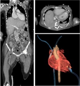
  • 0.23mm spatial resolution and Multi-Material Artifact Reduction to achieve high quality pan-scan with arms positioned along the body.


ASiR-V1
Reduce dose up to 82% as compared to FBP and Real-time reconstruction.

DoseWatch™
Dose management software solutions provide comprehensive data and analytics to help you:

  • Improve patient care by understanding each patient's exposure, identifying sources of variability, and improving protocol and exam delivery.
  • Meet regulatory and governing authority requirements for tracking and monitoring radiation dose.

Disclaimer

  1. In clinical practice, the use of ASiR, ASiR-V and Veo may reduce CT patient dose depending on the clinical task, patient size, anatomical location, and clinical practice. A consultation with a radiologist and a physician should be made to determine the appropriate dose to obtain diagnostic image quality for the particular clinical task. ASiR-V low contrast detectability (LCD), image noise, spatial resolution, and artifact were assessed using reference factory protocols comparing ASiR-V and FBP. The LCD measured in 0.625 mm slices and tested for both head and body modes using the MITA CT IQ Phantom (CCT183, The Phantom Laboratory), using model observer method. 


ASiR-V is available for Revolution EVO, HD and CT.

Post-processing

Post-processing


Real time reconstruction—Image Check
Image Check provides 340x340 matrix images for confirming Axial images in real time and tracking to up to 1800mm length with less than 1 sec delay. Reconstruction time is up to 55 fps.


Direct MPR
Direct MPR with Auto-Batch feature, affording automatic real-time direct reconstruction and transfer of fully corrected multiplanar images, in any plane.


One-Touch Protocol
One-Touch configurations allows you to customize the presentation of images according to the individual preferences of radiologists, so that the parameters of advanced treatment, the attributes of volume rendering, multiplanar reformatting, and the sizing of images are automatically applied when the series of patients starts.


 Advanced Visualization Workflow

  • Intuitive user Interface—Designed for infrequent users and experts and optimized visualization space.
  • Simplified workflows—Enhanced capture and recording of volumetric measurements.
  • Personalized workflow optimization—One-click access to customization list of high-frequency tools.
  • Dual monitor display option—AW Workstation and AW Server.


Chest Pain


  Scan setup and acquisition
  One-beat auto-gating scan setup and acquisition.


  Smart Phase
  Coronary specific best phase auto-selection.


  SnapShot Freeze
  Coronary specific motion correction.


Stroke


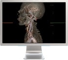
Autobone™ and Vessel-IQ Xpress: Head and neck CTA post-processing

• 1-click head and neck bone removal with intelligent vessel tracking.
• Comprehensive, 1-click stenosis measurement and plaque analysis.


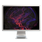
Dynamic 4D CTA: 4D whole brain CTA post-processing
• 1-click dynamic CT angiography analysis.
• Extracts CT angiography data from a CT Perfusion scan to visualize contrast flow.


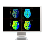
CT Perfusion 4D Neuro: Dynamic CTP post-processing
• 1-click CT Perfusion analysis with streamlined workflow for tissue classification.


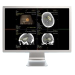
Stroke VCAR: Help to reduce "CT scan-to-report" time and "door-to-treatment" time.

Technologies

Patient Setup


Default patient positioning
Xtream Display provides workflow improvement by preset positioning (Default Patient Positioning) on the gantry display.


Special tables
Scan a whole-body trauma without having to move most of patients on the table with 2000mm long scannable range and 306Kg patient weight capacity.


One-stop scanning mode

  • Select the patient in one-touch.
  • Position patient hands-free.
  • Confirm and start scan.
  • All while at the gantry, without leaving the patient.


  1-click scan protocol setup
  Auto-load user configured trauma protocols.


  Smart patient centering
  Auto off-center direction and adjustments.


  Smart recon setup
  proactive DMPR ww/wl setup and image transfer.

Related Content

CT imaging in the Emergency Department - JB47984XX
RevolutionCT-Emergency-JB37939XE
JB00437CA October 2021