Not all products or features are available in all markets.

  • Change country/language

    Looks like you’re in {} — you’re on the {} site.

    Cancel

    Country and language selected

    You are being redirected to the {} site. Products and services availability vary by country. Do you wish to continue?

    Continue
    Cancel

MIMneuro™ brain PET/SPECT quantitative analysis software

Quantify and visualize PET and SPECT brain imaging with multi-tracer support, reference-database comparisons, advanced analysis methods and automated reporting.

icon
90% of the world's top 50 hospitals use MIM

Quantify Confidently with Centiloid*

MIMneuro includes quantitative analysis of amyloid plaque levels with the Centiloid scale at no additional cost. An automated workflow guides you through the process of generating and understanding results—you and your team have easy access to the data needed to make a confident assessment.

MIMNeuro-overview-easyanalysis

Brain PET and SPECT quantification across tracers and methods

  • Product Feature Icon
    Amyloid PET quantification with Centiloid scaling*
    Review amyloid PET with Centiloid, SUVR and Z-score outputs at no additional cost.
  • Product Feature Icon
    Template registration for amyloid PET analysis
    Register amyloid PET images to template space with MIM Workflows™ for quantitative review.
  • Product Feature Icon
    Multi-tracer PET and SPECT brain analysis
    Analyze supported PET and SPECT tracers and build a normals database for comparison.
  • Product Feature Icon
    Voxel-based, cluster and region-based analysis
    Choose quantitative methods including Z-score, SUVR and cortical surface projection views.
  • Product Feature Icon
    Brain image registration with BrainAlign™
    Match a patient brain scan to a normal-controls database for quantitative comparison.
Close details
Selected media

Quantitative neuroimaging methods and workflows

Explore MIMneuro capabilities for serial-exam comparison, brain image registration, atlas-based analysis and custom regions.

Serial PET and SPECT exam comparison

Compare baseline and follow-up brain exams with subtraction, normalization and cluster analysis workflows.
card1-half-width-mim-neuro-large-mr-pet-brain-career-fair

Visualize the difference

Compare baseline and activation PET and SPECT exams with a consistent process. Auto-normalization matches intensities so normal areas look similar on both exams. Cluster analysis highlights statistically significant differences and removes those that aren't statistically significant. Fuse the result to an MR or CT to localize the abnormality and share it with colleagues.

Custom brain regions for quantitative analysis

Create and save custom anatomical or combined brain regions for future quantitative analyses.
card2-half-width-mim-neuro-fdg-ssp-with-results

Advanced application

Researchers can take advantage of the same flexible and easy-to-use tools that are available to clinicians.


MIMneuro also provides the ability to create custom brain regions for analysis. Whether it is a new anatomical region or a combined region created by adding together several regions, custom brain regions can be saved for use in future quantitative analyses.

BrainAlign image registration

Match brain size, shape and orientation to a standard template for reference-database comparison.
card3-half-width-mim-neuro-mr-subtraction-with-results

Brain image registration for quantitative comparison

At the core of any neuro quantitative analysis software is the image registration method.

BrainAlign, a landmark-based deformation algorithm in MIMneuro, matches the size, shape, and orientation of a patient's brain scan to a database of normal controls for comparison.

Atlases for regional brain analysis

Use integrated atlases for regional statistics, SUVR calculations and normalization.
card4-half-width-mim-neuro-ptmr-fusion-normal

Single-brain atlas, probabilistic atlas for perfusion tracers and atlases generated for amyloid and DaTscan analysis.

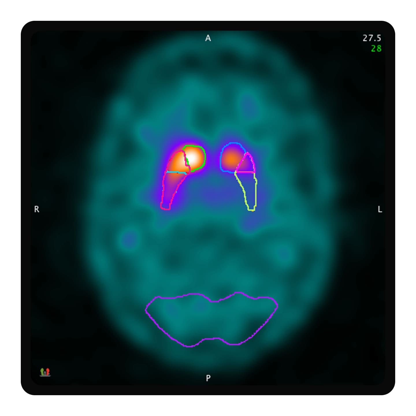
MIMneuro’s single-brain atlas is an anatomically complete atlas developed as a coordinated effort between several radiologists and a neuroanatomist. The probabilistic atlas for perfusion tracers is built from 10 individuals, allowing for increased anatomical sensitivity and specificity when performing region-based analysis. 

*Centiloid scaling in MIMneuro may not be available in your region. Contact your MIM Software representative for more information.

Get in touch

Have a question? We would love to hear from you.

JB04076XU November 2026
Contact us

We're ready to support you in your moments that matter.

We're sorry, a problem occurred with the form. Please try again.
Thank you for your inquiry

We will be in touch soon.

Something went wrong

We will check and get back.