NM/CT 860

The ideal balance of CT performance and clinical capability
At a Glance

Save time and steps

by remotely collaborating with a clinician mid-exam

Enhance productivity

with simplified workflows for complex procedures

Easy-to-use user interface

helps your department operate efficiently

Improve workflow

with automated transfer and archive features

NM/CT 860 is a SPECT/CT system designed for high-performance clinical environments

It combines the SPECT and productivity enhancements of the 800 Series with an optimal balance of CT technology. Technology with the thin-slice CT performance you need for all of your SPECT/CT protocols and the most common standalone CT exams, without overlapping with your other CT assets. NM/CT 860 is exactly what you need to continue to grow and strengthen the clinical value of nuclear medicine by making it more accessible in routine care.
Features

A SPECT/CT system for true discovery

You are committed to making true discovery even more accessible to patients. That's why we created NM/CT 860, to help you overcome any financial and operational challenges in your way, so you can continue to pursue true discovery.
TECHNOLOGY

Optimized for everyday imaging

In addition to the all-new SmartConsole, NM/CT 860 includes a new LEHRS (Low Energy High Resolution and Sensitivity) collimator, which can be combined with either SPECT Step & Shoot Continuous scanning mode or our Planar Clarity 2D processing to increase sensitivity. We paired these and other SPECT enhancements with our Revolution™ ACTs, an eight-slice CT system with fine resolution and contrast, as well as IQ Enhance, a reconstruction technology that scans at a faster helical pitch to cover more anatomy at similar image quality.

• Shorter photomultiplier tubes and lean front-end electronics reduce analog noise and improve performance

• Collimators optimized for high sensitivity, low septal penetration and high resolution

• Exceptional NEMA resolution for SPECT in a detector box that is 13 cm slimmer

• Evolution technology for up to a 50% reduction of scan time or patient dose1

• Enable a reduction of dose or scan times by up to 25% with the increased sensitivity of SwiftScan Planar and SwiftScan SPECT2

• Improved small lesion detectability with SwiftScan Planar and SwiftScan SPECT3

• ASiR™ technology can help you achieve your low-dose CT needs4
PRODUCTIVITY

Switch to an all-digital workflow with SmartConsole

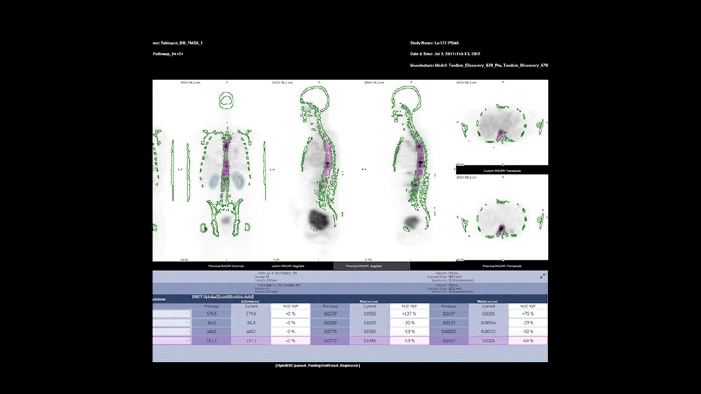
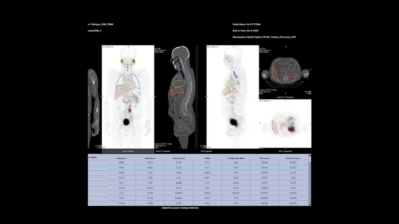
Experience a new productivity hub for hybrid imaging. By automating SPECT/CT reconstruction, SmartConsole simplifies the workflow for complex hybrid and quantitative protocols and allows technologists to review results directly at the scanner console. It also allows physicians to review scans remotely from mobile devices. They can modify processing settings and initiate study processing anytime, anywhere across a LAN or a WAN*. 

*Minimum hardware and software requirements apply

SmartConsole enables a noticeable improvement in productivity. Now, a physician can provide their input remotely and digitally. For example, physicians can review a whole-body bone scan directly on their tablet or other mobile device and define the scan range limits they need from wherever they are. The time saved with this switch to a digital workflow helps make it possible to complete exams in predictable time slots.
 
PRODUCTIVITY

One home for all of your molecular images

SmartConsole makes advanced procedures more accessible by transferring exams directly to the PACS or other pre-defined DICOM destinations with no operator intervention, even if SPECT/CT fusion is not supported, by exporting the studies in DICOM PET format. 

You can generate and send an additional dataset in PET DICOM format to enable quantitative SPECT results and SPECT SUV (Standard Uptake Value) on any PET DICOM compliant workstation or PACS. Referring physicians can review and evaluate your SPECT/CT exams with the same quality of information they have come to expect from your PET/CT, as adjunct information to the original SPECT/CT images.

Capability

The right mix of SPECT and CT capability
Until now, many advanced hybrid and quantitative protocols have been limited to research institutions with the extra resources needed to provide complex protocols. Through SmartConsole and Xeleris™, NM/CT 860 makes these protocols available in routine settings by reducing the resources needed to perform them.

A simpler way to bring everything together

Q.Volumetrix MI is an Xeleris 4 DR application that integrates SUV measurements directly into the volume viewer. It allows you to see both the image and the SUV simultaneously instead of having to switch back and forth between two separate applications. Then, with SmartConsole, your quantitative SPECT/CT studies are automatically transferred directly to the PACS or other pre-defined DICOM destination. These resource-saving improvements allow you to expand your services without breaking your schedule.

Stay on top of the latest trends in imaging

Grow your referral volume by offering surgical planning procedures.


A CT that stands out when it stands alone

Revolution ACTs gives you the thin-slice performance you need for all of your SPECT/CT exams and standalone CT capability for the most common overflow CT studies. Not only that, but you get the added time savings of being able to acquire a diagnostic CT exam at the same time as a SPECT/CT exam, which helps improve the patient experience. In addition, Revolution ACTs has Smart Dose technology referring physicians expect with every exam.

Features

Additional benefits provided by NM/CT 860

Streamline access to SPECT/CT

and quantitative SPECT/CT studies

Optimize image quality

post-exam with retrospective processing features

Reduce cost and save space

by sharing collimators with existing 600 Series and 800 Series systems

Future-ready

Modular design enables option to purchase SPECT advancements and access to image quality enhancements
DISCLAIMERS
1. In clinical practice, Evolution options1a (Evolution for Bone, Evolution for Cardiac, Evolution for Bone Planar) and Evolution Toolkit1b are recommended for use following consultation of a Nuclear Medicine physician, physicist and/or application specialist to determine the appropriate dose or scan time reduction to obtain diagnostic image quality for a particular clinical task, depending on the protocol adopted by the clinical site.

1a. Evolution Options - Evolution claims are supported by simulation of count statistics using default factory protocols and imaging of 99mTc based radiotracers with LEHR collimator on anthropomorphic phantom or realistic NCAT – SIMSET phantom followed by quantitative and qualitative images comparison.

1b. Evolution Toolkit - Evolution Toolkit claims are supported by simulation of full count statistics using lesion simulation phantom images based on various radiotracers and collimators and by   showing that SPECT image quality reconstructed with Evolution Toolkit provide equivalent clinical information but have better signal-to-noise, contrast, and lesion resolution compared to the   images reconstructed with FBP / OSEM.

2. Compared to LEHR collimator, with Step & Shoot scan mode (for SPECT) / without Clarity 2D (for Planar). As demonstrated in phantom testing using a bone scan protocol, Evolution processing (for SPECT), and a model observer. Because model observer results may not always match those from a human reader, the actual time/dose reduction depends on the clinical task, patient size, anatomical location and clinical practice. A radiologist should determine the appropriate scan time/dose for the particular clinical task.

3. As demonstrated in phantom testing using a model observer. For SPECT, compared to using the LEHR Collimator and a SPECT Step & Shoot acquisition. For Planar, compared to using LEHR without Clarity 2D.

4. In clinical practice, the use of ASiR or VISR may reduce CT patient dose depending on the clinical task, patient size, anatomical location and clinical practice. A consultation with a radiologist and a physicist should be made to determine the appropriate dose to obtain diagnostic image quality for the particular clinical task.

Have a question? We would love to hear from you.

JB17578XX