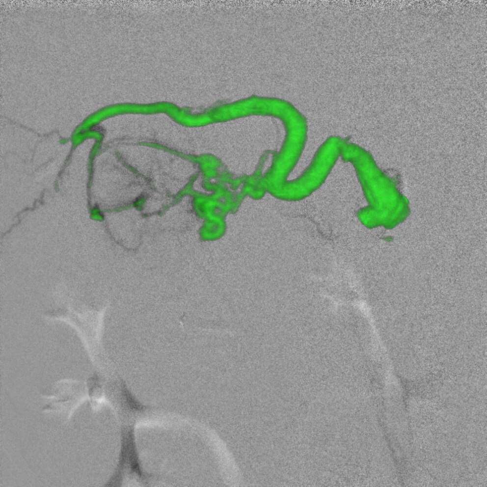
Liver embolization
To deliver therapeutic material to tumors during trans-arterial chemoembolization therapy, it is essential to identify the liver vessels accurately. But the liver's complex vasculature can make precise identification of tumor-feeding vessels in 2D and 3D images a challenge, often requiring significant time, radiation, and contrast media.
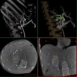
Plan
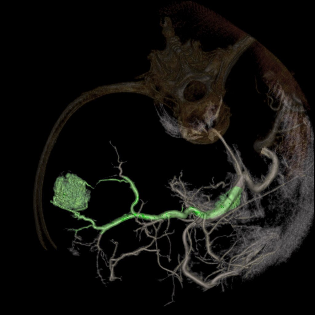
FlightPlan for Liver helps you plan your liver embolization procedure. It automatically highlights vessels traveling from the catheter tip to the vicinity of a liver hypervascular lesion.
Guide
Once ready, you can send the FlightPlan for Liver 3D model to Innova Vision with a single click and use it as a 3D roadmap to guide catheters across tortuous vessels and bifurcations, helping you perform the embolization with confidence.
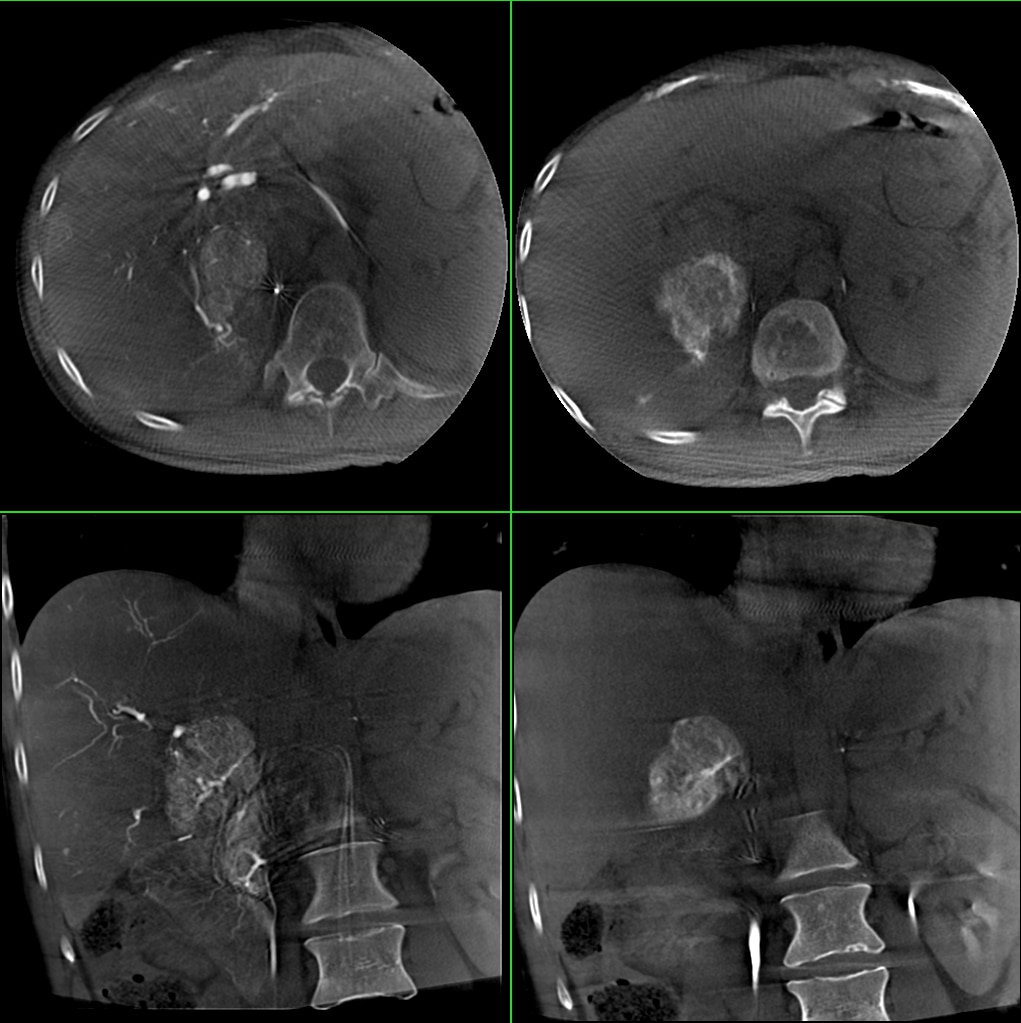
Assess
A post-operative Innova CT helps you determine the success of the embolization.
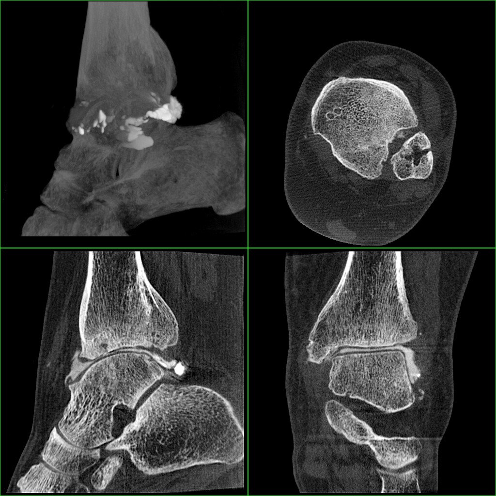
Needle procedures
Performing needle procedures in the interventional suite frees up your CT system and provides better access to the patient. However, under fluoroscopic guidance, it may be challenging and time-consuming to find the right entry point and advance the needle while avoiding critical structures.
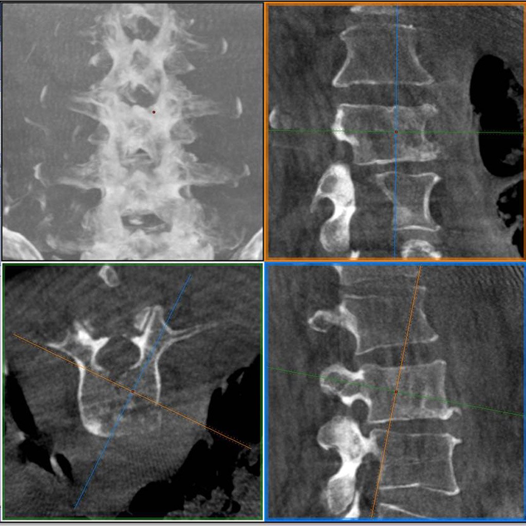
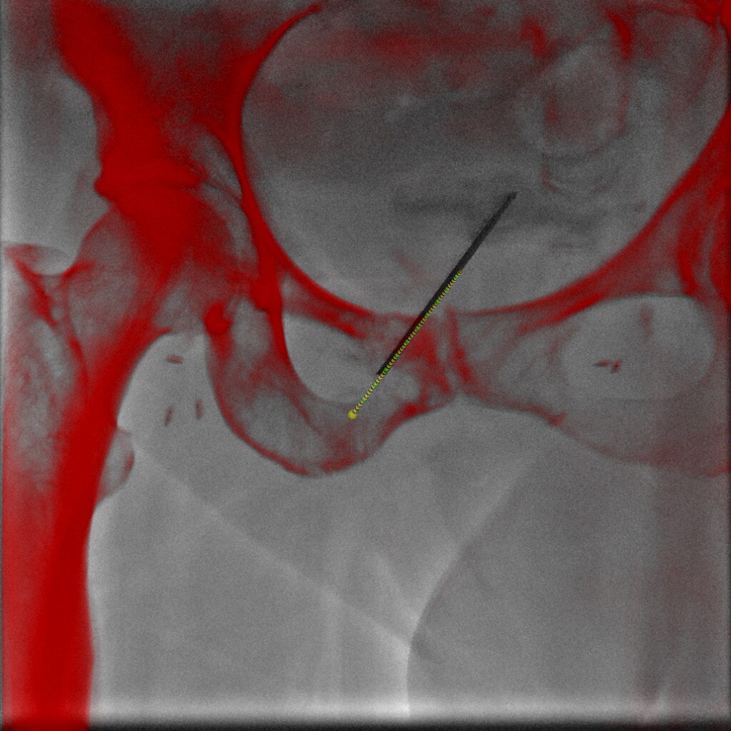
Plan
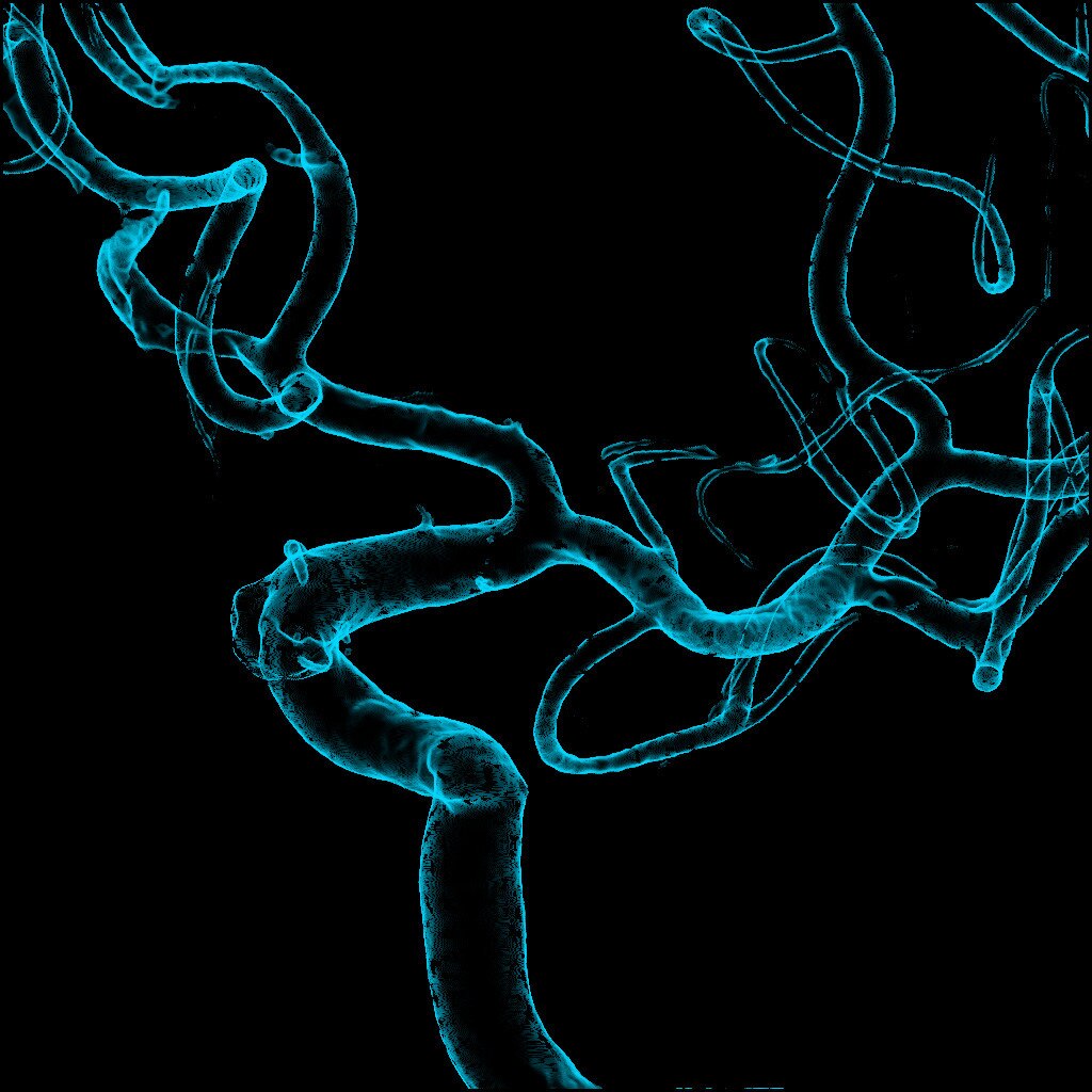
With Volume Viewer and Trajectory Planning, you can plan the procedure using outstanding 3D information and determine the optimal skin entry points and needle paths.
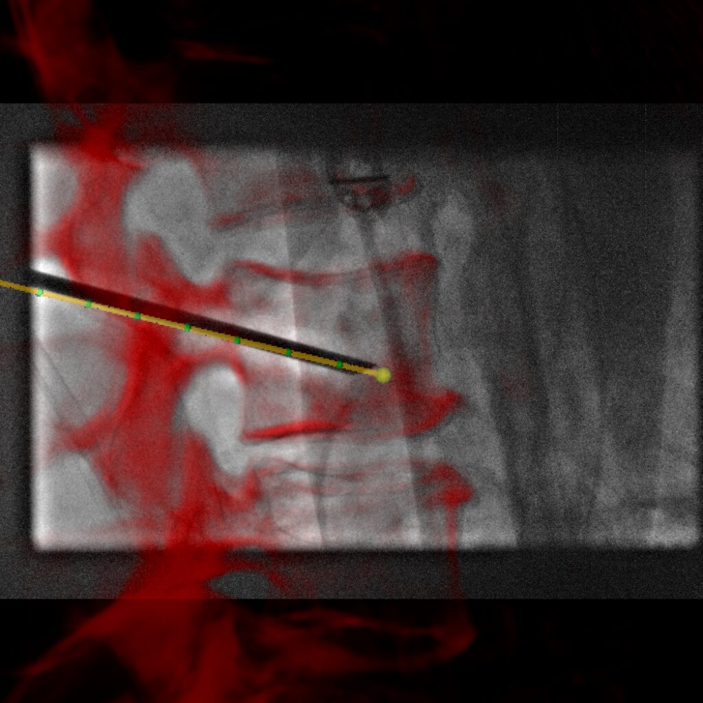
Guide
With Innova TrackVision, you can guide your needle along the trajectory that will follow the C-arm angulation and table movement. Plus, a dedicated algorithm overlays the bone anatomy and helps you correct for even small patient motion, helping to facilitate accurate needle trajectory registration at any time during your procedure.
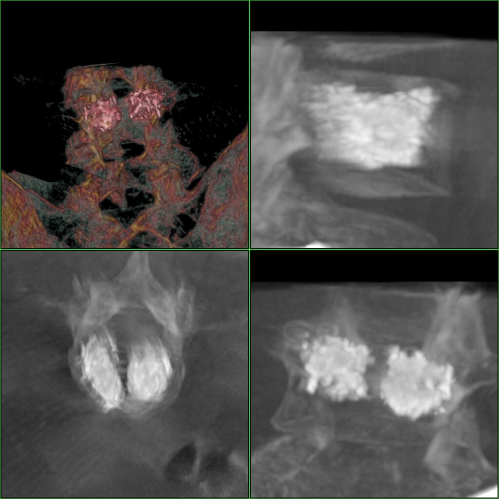
Assess
At the end of the procedure, when the cement has been injected, an Innova CT acquisition will help you see the cement implantation in 3D and thus confirm procedure completion.
Portal Vein Embolization
The objective is to embolize only the branches that correspond to the part of the liver that will be resected. The portal vein has numerous anatomical variations, with many different branches that can be difficult to distinguish one from another on 2D fluoroscopic images. To do this with confidence, you must understand the exact position of the catheter in the anatomy and control the embolization phase in real time.
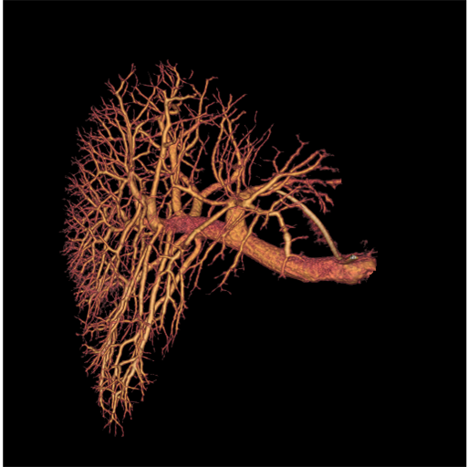
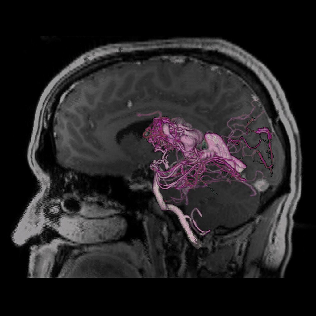
Plan
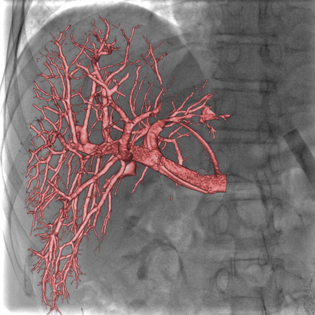
With Innova 3D, you can depict the full liver portal system and select the branches to be embolized without being bothered with vessel tortuosity and superimposition issues.
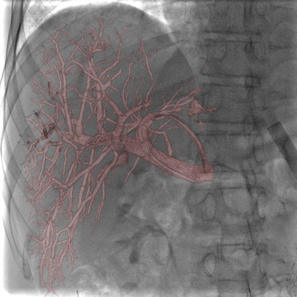
Guide
Using Innova Vision, you can fuse the 3D information on live fluoroscopy in the lab with real-time adjustment of the C-arm angulations and table position, offering virtual support to navigate in the anatomy and control the embolic agent material injection.
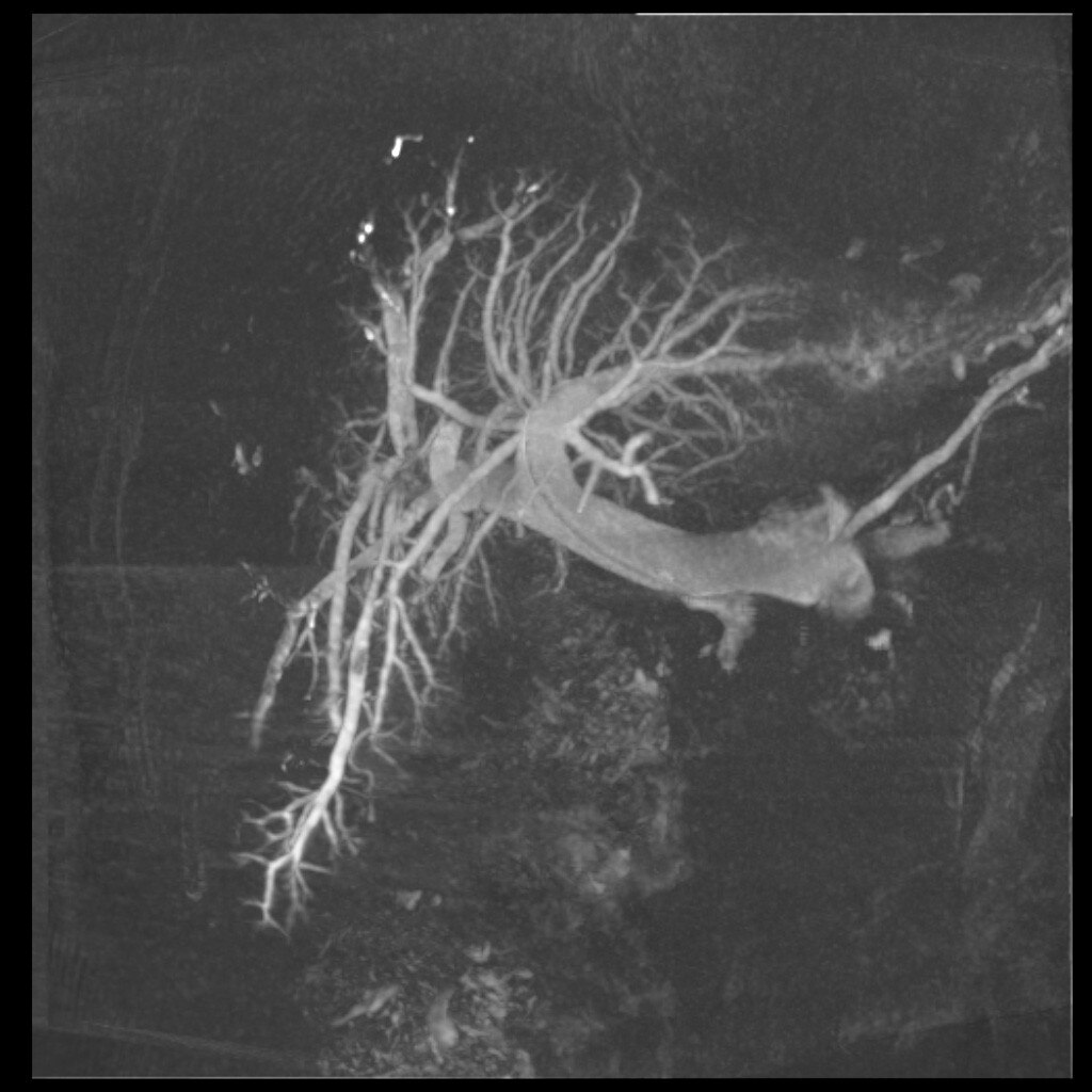
Assess
After the procedure, you can use an Innova Subtracted 3D acquisition to visualize embolic material uptake in the branches embolized allowing you to determine whether the patient needs additional branches embolized.