As a provider in a high-performing clinical environment, you constantly
look for opportunities to adopt the latest in imaging technologies, like
spectral imaging. However, you can't afford to adopt this technology if
it has limited utility or doesn't fit into the processes you already have in
place. We addressed this challenge with GSI Pro.
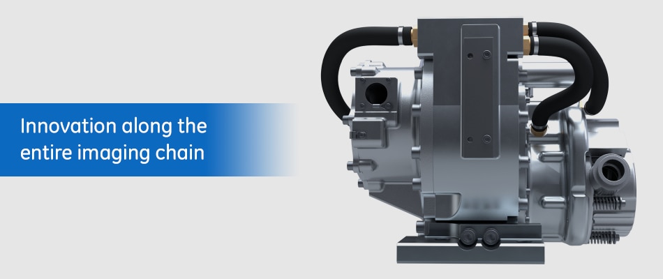
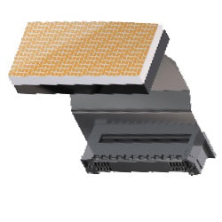
GSI is our proven spectral imaging application that uses the
Gemstone detector and rapid kV switching to acquire dual energy
samples from a single source. By rapidly switching between two
different kV energies at a rate of up to 0.25 ms with sub-millimeter Z-axis
registration, its temporal registration is over 165 times faster than
other dual energy technologies. And the advantage of its single source
architecture is the ability to generate material decomposition images
over the full 50 cm field of view.
We addressed and improved the GSI experience to seamlessly
integrate with AW applications and to significantly reduce
reconstruction times. It's a breakthrough in spectral CT technology
that effortlessly processes gigabytes of data at a time, making the
clinical benefits of GSI routinely accessible.
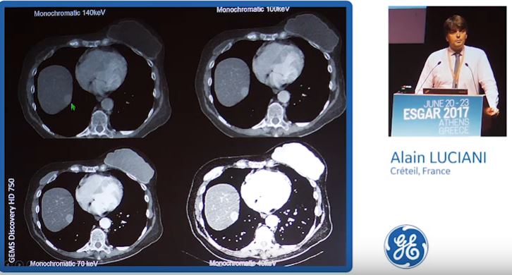
The clinical benefits of GSI Pro include up to a 50 percent
improvement in beam-hardening artifact due to bone, metal and other
high-contrast materials such as iodine. It also has the ability to deliver
non-contrast-like images by subtracting detected iodine from an image. By
incorporating the latest in iterative reconstruction technology, ASiR-VTM,2
enables dose neutrality, lower image noise and improved low contrast
detectability for patients of any size.
Improve clinical
outcomes

With over 300 publications on
GSI, example findings* by the
academic community include:
liver lesion detection by 17% and
kidney lesion characterization by
12%, reducing the need for
unnecessary follow-upsa,b,
reduce contrast by at least 50%,
benefitting patients with renal
functionc,d,e and 6 times
reduction in non-interpretable
scans with GSI MARf,g. (see GSIInfographic)
*The example findings cited are limited to the referenced studies only and may not be broadly applicable to your clinical practice.
References
a. Marin, D., et. al. "Characterization of Small Focal Renal Lesions: Diagnostic Accuracy with Single-Phase Contrast-enhanced Dual-Energy CT with Material Attenuation Analysis Compared with Conventional Attenuation Measurements." Radiology. 284, no. 3 (2017).
b. Liu, Qi-Yu, et. al. "Application of gemstone spectral imaging for efficacy evaluation in hepatocellular carcinoma after transarterial chemoembolization." World Journal of Gastroenterology 22, no. 11 (2016): 3242.
c. White Paper of the Society of Computed Body Tomography and Magnetic Resonance on Dual-Energy CT, Part 2: Radiation Dose and Iodine Sensitivity; Part 3:Vascular, Cardiac, Pulmonary and Musculoskeletal Applications; Part 4: Abdominal and Pelvic Applications. J Comput Assist Tomogr (2016).
d. Dong, Jian, et al. "Low-contrast agent dose dual-energy CT monochromatic imaging in pulmonary angiography versus routine CT." J of computer assisted tomography 37, no. 4 (2013): 618-625.
e. Shuman, William P., et. al. "Prospective comparison of dual-energy CT aortography using 70% reduced iodine dose versus single-energy CT aortography using standard iodine dose in the same patient." Abdominal Radiology 42, no. 3 (2017): 759-765.
f. Reynoso, Exequiel, et. al. "Periprosthetic Artifact Reduction Using Virtual Monochromatic Imaging Derived From Gemstone Dual-Energy Computed Tomography and Dedicated Software." J Comput Assist Tomogr . 2016; 40 (4): 649-657.
g. Pessis, Eric, et. al. "Virtual Monochromatic Spectral Imaging with Fast Kilovoltage Switching: Reduction of Metal Artifacts at CT" RadioGraphics 2013; 33:573-583.