• Change country/language

    Looks like you’re in {} — you’re on the {} site.

    Cancel

    Country and language selected

    You are being redirected to the {} site. Products and services availability vary by country. Do you wish to continue?

    Continue
    Cancel

Access to spectral CT information without impacting your clinical workflow


Leverage the power of Spectral Imaging with 3D advanced visualization capabilities


Gemstone Spectral Imaging (GSI) has the exciting potential to be used as a routine scan acquisition mode offering additional anatomical and functional information to help expedite and assist in your CT diagnosis: Expand your spectrum of clinical capabilities with GSI without impacting your clinical workflow.
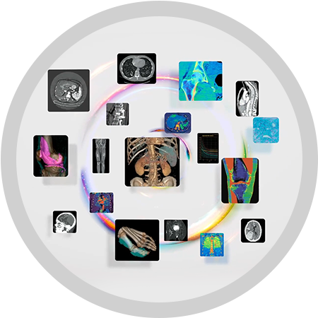
GSI Viewer

Aids in confident diagnosis with easier workflow

The GSI Viewer is the primary method of viewing and manipulating spectral images. It allows for the review of monochromatic energy images at user selectable energy levels, detailed analysis using material decomposed images (such as water-iodine, water calcium, etc.), and complementary information using the Effective-Z (provides an estimate of the protons, Z number, in a voxel) images. GSI Viewer leverages the power of Spectral Imaging with 3D segmentation and advanced visualization capabilities.

GSI Viewer

Expand your spectrum of clinical capabilities with spectral imaging

Monochromatic review

Review the monochromatic images in multi-planar, curved and lumen reformats and in 3D VR mode. Interactively change the monochromatic energy levels to select the best energy level for the exam being reviewed.
AW-GSI-Viewer-Monochromatic-review

Image Overlay

Overlay material density or Effective Z information on top of the monochromatic images.
AW-GSI-Viewer-Image-Overlay

Material Suppressed Iodine/Virtual Unenhanced (VUE)

Suppress iodine to assist in characterizing or identifying lesions/pathology.
AW-GSI-Viewer-Material-Suppressed-Iodine

Material Density Analysis

Visually see how the GSI data is segregated amongst a material density pair, e.g. water and iodine
AW-GSI-Viewer-Material-Density-Analysis

Plot Analysis

Displays ROI’s as graphical plots (histogram, spectral HU curve and normalized spectral HU curves) to visualize how a selected region behaves at different energy settings.
AW-GSI-Viewer-Plot-Analysis

Liver Fat

Liver Fat Quantification. Evaluate hepatic steatosis with liver fat percentage measurements.
AW-GSI-Viewer-Liver-Fat

GSI Pulmonary Perfusion²

One click protocol for relative iodine distribution to assist in identification of relative hypo-perfused territories.
AW-GSI-Viewer-GSI-Pulmonary-Perfusion

GSI Gout

Fully automated workflow for material decomposition and color overlay to assist in gout characterization.
AW-GSI-Viewer-GSI-Gout

Leverage the power of Spectral Imaging with 3D advanced visualization capabilities

GSI Gout Protocol

Fully automated workflow for material decomposition and color overlay to assist in gout characterization:


  • Automated generation of 2D and 3D gout overlay
  • Automated export of 2D and 3D gout overlay images to the patient browser
S-Curve-GSI-Gout-Protocol

GSI Pulmonary Perfusion²

Dedicated one-click protocol protocol to assist in identification of relative hypo-perfused territories:


  • Automatic relative hypo-perfused region display
  • Automated export of 2D and 3D gout overlay images to the patient browser
  • Volumes reported for total lung and relative hypo-perfusion volumes
  • Succinctly communicate volumes and relevant pictures to referring physicians
S-Curve-GSI-Pulmonary-Perfusion

GSI Fat

Dedicated GSI fat capability to quantify fat in the liver:


  • Evaluate hepatic steatosis with liver fat percentage measurements
  • GSI with material separation provides ability to measure approximate liver fat percentages
  • Liver fat measurements compatible with non contrast¹ and multi-phase contrast acquisitions
s-curve-gsi-fat-desktop

FOOTNOTES

  1. GSI Liver Fat ROI’s can be done on non-contrast or any phase of contrast enhancement.
  2. GSI Pulmonary Perfusion protocol is an optional add-on protocol to Thoracic VCAR application.
Contact Us

We're ready to support you in your moments that matter. For service assistance - +1 800-437-1171

Thank you!

We received your request for assistance.

A GE Healthcare specialist will be reaching out shortly.

Something went wrong

We will check and get back.

JB01114AU May 2023