1. In clinical practice, Evolution options 1a (Evolution for Bone, Evolution for Cardiac, Evolution for Bone Planar) and Evolution Toolkit 1b are recommended for use following consultation of a Nuclear Medicine physician, physicist and/or application specialist to determine the appropriate dose or scan time reduction to obtain diagnostic image quality for a particular clinical task, depending on the protocol adopted by the clinical site.
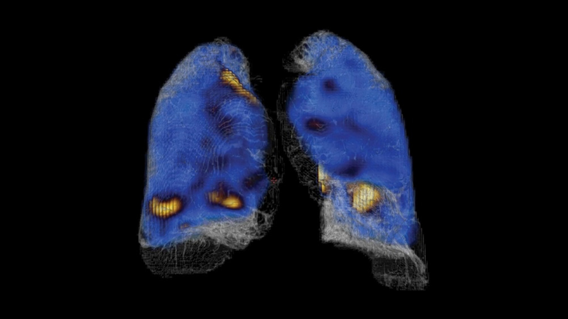
1a. Evolution Options - Evolution claims are supported by simulation of count statistics using default factory protocols and imaging of 99mTc based radiotracers with LEHR collimator on anthropomorphic phantom or realistic NCAT – SIMSET phantom followed by quantitative and qualitative images comparison.
1b. Evolution Toolkit - Evolution Toolkit claims are supported by simulation of full count statistics using lesion simulation phantom images based on various radiotracers and collimators and by showing that SPECT image quality reconstructed with Evolution Toolkit provide equivalent clinical information but have better signal-to-noise, contrast, and lesion resolution compared to the images reconstructed with FBP / OSEM.
2. Compared to LEHR collimator, with Step & Shoot scan mode (for SPECT) / without Clarity 2D (for Planar). As demonstrated in phantom testing using a bone scan protocol, Evolution processing (for SPECT), and a model observer. Because model observer results may not always match those from a human reader, the actual time/dose reduction depends on the clinical task, patient size, anatomical location and clinical practice. A radiologist should determine the appropriate scan time/dose for the particular clinical task.
3. As demonstrated in phantom testing using a model observer. For SPECT, compared to using the LEHR Collimator and a SPECT Step & Shoot acquisition. For Planar, compared to using LEHR without Clarity 2D.
4. In clinical practice, the use of ASiR or VISR may reduce CT patient dose depending on the clinical task, patient size, anatomical location and clinical practice. A consultation with a radiologist and a physicist should be made to determine the appropriate dose to obtain diagnostic image quality for the particular clinical task.
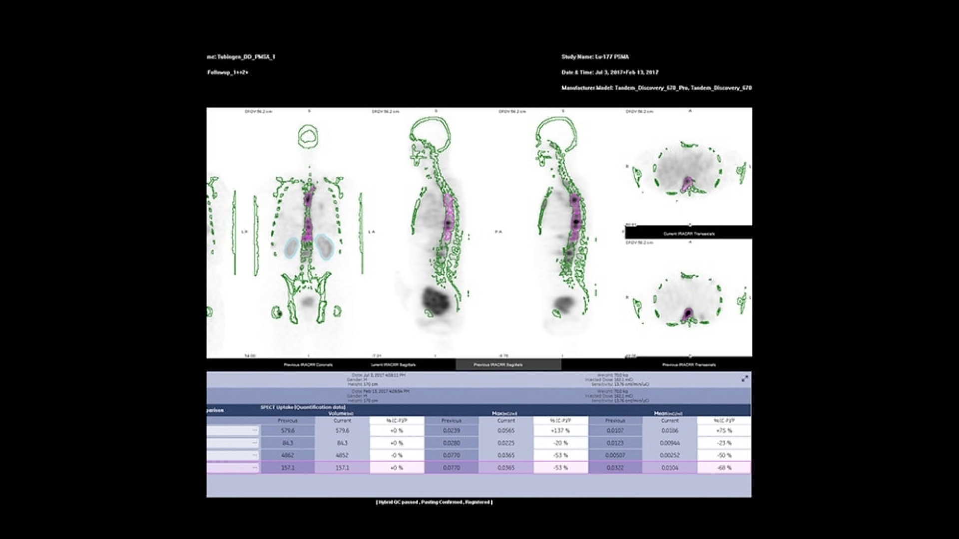
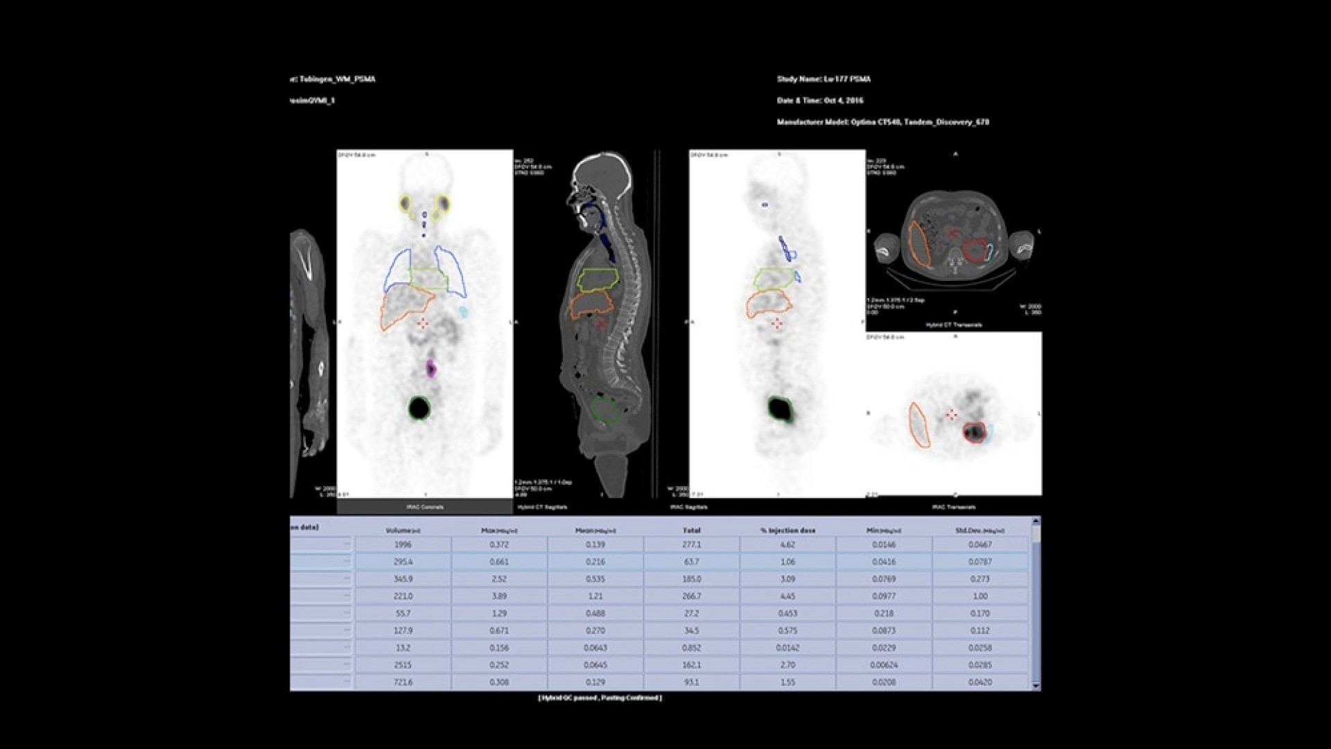
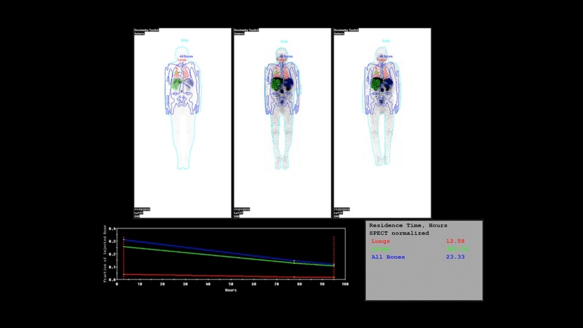
5. Compared to using Xeleris 4 DR Dosimetry Toolkit with dose calculations done manually or with external software. As demonstrated in an evaluation at two sites consisting of 14 SPECT/CT cases by four physicists, where each case was processed by one of the four physicists. Results may