Don't have an account?
StarGuide*
Introducing StarGuide, our most advanced SPECT/CT featuring advanced Cadmium Zinc Telluride (CZT) technology.



Explore new protocols by visualizing multiple tracers simultaneously

Search for new insights with 3D Dynamic Acquisition¹

Research existing tracers to help expand their clinical applications

Support the development of new tracers

StarGuide provides accurate quantitation to help clinicians make personalized care decisions and treatment response assessments that are at the heart of Theranostics by striking the right balance of energy resolution and spatial resolution. Each of StarGuide’s 12 CZT Digital Focus Detectors supports a wide energy range capable of imaging the dual energy peaks of tracers used in Theranostics, such as the 113 keV and 208 keV emission peaks of 177Lu used to diagnose and evaluate treatment response for prostate cancer². StarGuide is optimized for 177Lu imaging, enabled by its improved energy resolution, system sensitivity, and system planar resolution for 177Lu high and low energy peaks as compared to a NaI system³. This enables 177Lu imaging without the need for collimator exchange — typically needed in dual head cameras — simplifying day-to-day clinical workflow and patient scheduling.

GE Healthcare is at the forefront of molecular imaging, and has been since we began development of the first commercially available SPECT/CT scanner in 1999 and PET/CT in 2000. To maintain our leadership position, we have established the CZT Center of Excellence in Rehovot, Israel, where we continuously develop advanced, proprietary CZT technology found exclusively in GE Healthcare’s Nuclear Imaging products. Our commitment to meeting our customers’ nuclear medicine needs extends to the manufacture and distribution of radiopharmaceutical products as well. Through GE Healthcare’s Pharmaceutical Diagnostics (PDx) division, we’re the only medical equipment manufacturer with the ability to develop and supply highly specialized pharmaceuticals to clinical and research institutions throughout the world.
StarGuide’s Optical Scout feature represents a genuine breakthrough in wholebody scanning and charts the path toward high quality clinical images. Located within its 80 cm bore, Optical Scout performs an optical scan that creates an accurate topographical map of the patient, quickly and accurately scanning body contours to assure fast and safe transition from one imaging position to the next.
After processing the contour data, StarGuide’s detectors and table are automatically positioned in close patient proximity for each bed position before beginning the clinical scan. With patient safety of paramount importance, each detector also contains a pressure sensitive cover, stopping motion if it is triggered.
Innovative design is nothing new to the engineers at GE Healthcare. When we first began the design for StarGuide, we put great emphasis on performance standards for SPECT/CT imaging with the goal of helping improve patient outcomes. We are gratified these efforts have resulted in StarGuide winning the prestigious 2021 Red Dot Award for product design.

StarGuide’s Digital Focus Detectors are designed to propel SPECT imaging to new heights in performance by maximizing the amount of information to help you gain true insight into the patient condition. After optimizing the detector and bed positions from the Optical Scout data, StarGuide’s constellation of 12 slim Digital Focus Detectors orbit the body as closely as possible, and from all necessary angles, to scan the target area — and not the air surrounding the patient. The result is a high-resolution scan and minimization of patient time on the camera. CZT detector technology features a higher count rate over conventional detector technologies. StarGuide’s intrinsic count rate is greater than 700 kcnts/second with no detector saturation or resulting dead time.


The CZT material at the heart of each Digital Focus Detector provides an inherently high contrast-to-noise ratio. Its 2.46 mm² pixel size and 8 mm spatial resolution⁴ deliver the clarity you need to detect, locate, and quantify diseases at an early stage – helping to open the door to more effective treatments and improved patient outcomes. StarGuide delivers a remarkable 5.9% energy resolution when scanning ⁹⁹ᵐTc, and <5% energy resolution when scanning 177Lu at its 208 keV peak. With such superb energy resolution, StarGuide gives you the ability to effectively differentiate between simultaneously acquired isotopes with smaller differences in peak energies.
Image a wide energy range
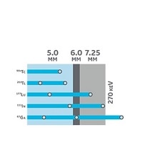
The ability to image both low and medium energy isotopes in a single scan with exceptional levels of spatial and energy resolution is what makes StarGuide a great tool for both clinical and research environments. The proprietary design of our 7.25 mm thick CZT semiconductor crystals makes it possible to image a wide range of isotopes
with energies up to 270 keV.
Take a closer look where needed

Focused imaging gives you the ability to target a specific volume of interest (VOI) within the patient’s body and improves image quality within that VOI as the Digital Focus Detectors image that volume. StarGuide alters the detector sweep to spend more time focusing on the designated area to produce high-quality, data-rich imagery
with no increase in scan time.
Analyze flow in greater detail
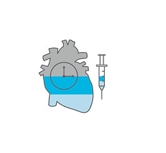
StarGuide’s ring of Digital Focus Detectors enables Dynamic SPECT acquisitions1 with high temporal resolution as fast as 5 seconds. Fast temporal resolution is an important factor in imaging bolus injection and flow measurements.
Personalized patient exposure
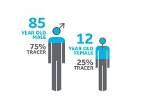
StarGuide’s CZT technology is pivotal to minimizing the patient’s exposure to SPECT’s radioactive processes because its higher system planar sensitivity and volume sensitivity is an important factor in NM imaging for enabling dose
and time reduction5..
Dual Channel collimation
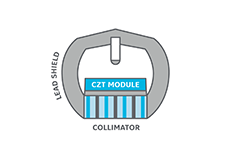
StarGuide’s exclusive Dual Channel collimator enables a balance between sensitivity and resolution across low and medium energy ranges. Its design combines shorter septa and tungsten construction to deliver a remarkable 8.0 mm NEMA planar resolution and 4.5 mm Central SPECT resolution with scatter. The improved SPECT resolution enables sharp images for better visualization of fine anatomical details, which can help physicians to detect diseases
at an earlier stage6.

StarGuide’s image processing engine is designed to help physicians advance from analog 2D scanning to digital 3D scanning in their pursuit of true insight into the patient condition. The 12 Digital Focus Detectors scanning in 3D generate exponentially more data than previous 2D scans, significantly increasing the processing power of StarGuide’s image processing engine to efficiently calculate the thousands of additional vectors, variable bed positions, and other factors. The result is a high-quality image. Innovative physicians looking to navigate molecular imaging’s unexplored regions will appreciate that StarGuide’s image processing engine offers highly customizable reconstruction capabilities in addition to a robust set of default protocols to steer their pursuits in any direction they choose.
The Q.Clear reconstruction algorithm represents a significant step forward for use in SPECT imaging, including in the assessment of treatment response in addition to the diagnosis and staging of disease. This advancement is made possible by suppressing noise to provide outstanding quantitation accuracy without sacrificing StarGuide’s ability to accurately determine lesion size and volume.
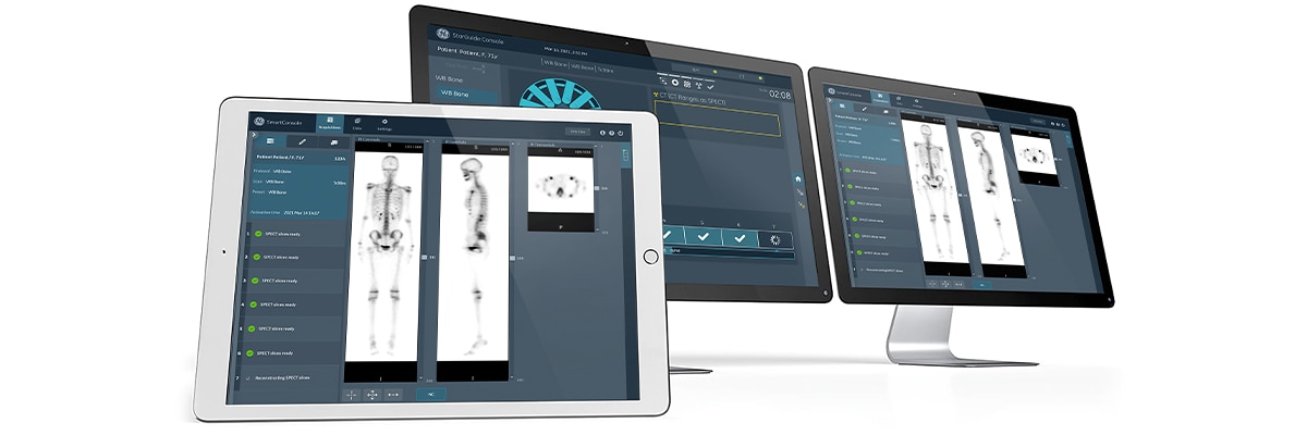
SmartConsole is our latest network-capable image processing sub-system for Nuclear Medicine that enables you to automate SPECT/CT reconstruction, simplify complex hybrid imaging and quantitative protocols, and generate high-quality hybrid images so you can make informed decisions that lead to quantifiable results. SmartConsole is optimized to host our latest reconstruction algorithms and applications, plus it helps you seamlessly clarify and share patient data. SmartConsole includes FAME post-processing, which integrates the CT anatomical bone information into the SPECT bone image processing. When used on StarGuide images, the lesion detectability is improved, which helps increase diagnostic confidence⁷.

Swift Plan workflow is designed to simplify clinician interactions with a scanner as fast and sophisticated as StarGuide. By leveraging proven GE Healthcare innovations like its interactive table ruler and our advanced automation robotics, then combining them with new innovations like our intuitive bedside display and Optical Scout, Swift Plan quickly becomes an extremely capable and user-friendly productivity enhancer. Swift Plan with SmartConsole automates SPECT/CT reconstruction, simplifies complex hybrid and quantitative protocols, optimizes image quality with retrospective processing features, reduces errors, allows operators to see images at the scanner console, and enables physicians to review scans remotely. Even with a long list of capabilities, the learning curve for Swift Plan is remarkably short.


Reliable scheduling

Replace planar exams with the predictable, repeatable scan times of 3D-only protocols which may minimize the need for additional views, reduce scheduling overruns and shorten patient wait times.
Scanning comfort

StarGuide’s Swift Plan minimizes the time the patient spends motionless on the camera, helping lower the stress for patients with certain anxieties.
Higher Sensitivity⁵

Leverage StarGuide’s higher system planar sensitivity and volume sensitivity, an important factor in NM imaging for enabling dose and time reduction.
Fewer rescans

Optical Scout, Swift Plan, StarGuide’s image processing engine, and Focused Imaging are important factors in helping reduce the need for inconvenient and anxiety-inducing rescans.
Rapid Contouring

Optical Scout’s contour scans help minimize the likelihood of detector contact while enabling close
patient proximity.
High-resolution images

Exceptional image quality and the enormous amount of high-resolution data that StarGuide generates with each scan give physicians a clear image.
Multiple tracers

StarGuide has the ability to simultaneously image multiple tracers in a single scan that may otherwise involve multiple patient visits and, in relevant cases, multiple patient sedations. This inherent temporal registration helps increase the confidence in
differential diagnosis.


Xeleris V*

MyoSPECT