Hesabınız yok mu?
Innova™ IGS 540 girişimsel onkoloji prosedürleri için en büyük görüntüleme alanlarından birini içeren, zemine monte bir sistemdir.


With its 41 cm (16.1 in) square detector, Innova IGS 540 provides extensive body coverage for peripheral and abdominal procedures.
The Innova IGS 540 was designed from the ground up to provide the image clarity you need while helping you keep dose as low as possible. It includes features like Dose Personalization1, which gives you the tools to choose from up to five2 automatic exposure preferences for your system. You can also modify any of these preferences in any clinical protocol, to enable multi-procedure, multi-user customization, and thus support well-informed decisions.
Powered with dedicated advanced applications, you can plan, guide and assess procedures with confidence.
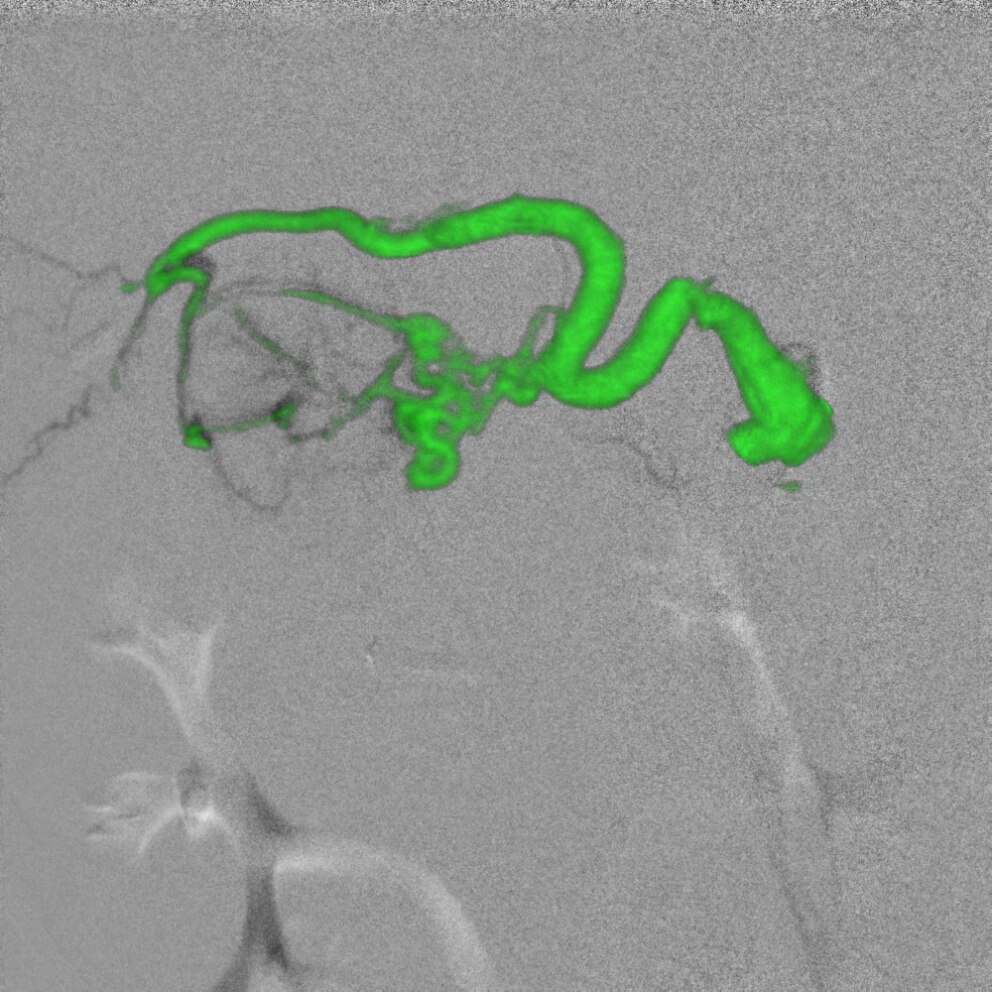
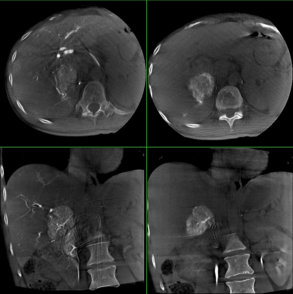
FlightPlan for Liver helps you plan your liver embolization in three intuitive steps and automatically highlight vessels traveling from the catheter tip to the hyper-vascular tumor.
Innova TrackVision helps you can plan and guide needle procedures by identifying one or multiple needle trajectories for your case and overlay these virtual trajectories over the live fluoroscopic images - so you know where you are in delicate
3-axis C-arm makes complex angulations easy to achieve.
Maximum positioning flexibility and excellent patient access for coverage from groin to head.
Comfortable access to the head of the patient with the offset C-arm and L-positioner.
Easy transition from AP to lateral using flexible offset C-arm and L-positioner.
Follow the contrast bolus in real time using variable panning speed control with an easy workflow.
3D model moves in synchrony with the gantry angulations.
Simple, centralized, automated control of system functions from the tableside.
Navigate at tableside in the anatomy on pre-acquired or per-operative datasets.
Easy 200° rotational spin with automatic volume reconstruction.
Intuitive 3-step liver embolization planning at the beginning of the procedure.
Helps you precisely plan needle trajectories, progress with confidence down the planned trajectory overlaid on live fluoroscopy, and visualize any deviations from the path.
Innova Vision
The Innova IGS 540 with its 41 x 41 cm (16.1 in) square flat-panel digital detector boasts one of the largest fields of view for vascular and interventional imaging. It can cover large anatomies, such as both legs simultaneously, designed for fewer runs than smaller detectors, enabling efficient use of contrast and dose.

The proprietary angiography flat-panel detector offers one of the industry’s highest ratings for Detective Quantum Efficiency (DQE), a parameter internationally acknowledged as an index of detector performance in contrast- and dose-limited imaging performed in clinical studies. High DQE enables better-quality images at the same dose, or the same quality image at a lower dose.

GE designs systems from the ground up with the tried and trusted GE imaging chain, optimized to provide the image clarity you need while keeping dose as low as possible. Our dose-reduction features empower you to easily optimize and personalize dose settings from the tableside, while maintaining clinical details you need to make well-informed decisions. But improving dose management takes a strategy – what we call the GE Blueprint. It includes low-dose imaging technologies for minimally invasive procedures, but also considers the people, culture and processes around them.

Control your system and images with integrated, intuitive tableside controls. With simple menus, the Central touchscreen lets you control most system functions, configure the system, modify imaging parameters, control your large display monitor, and manipulate advanced applications – all at the tableside. The comfortable, easy-to-grasp control knob makes it easy to pan the table, position the gantry, and perform procedures.

The full-color 142 cm (56 in) diagonal 8 megapixel medical-grade large display lets you view multiple images from multiple sources. Get up to 120 customized layouts, easily changeable at tableside with the Central touchscreen. Zoom in comfortably without loss of detail or pixilation to get the clinical focus you need.

Over 20 advanced applications to help you plan, guide and assess complex procedures
GE Healthcare offers a full suite of advanced applications designed to help improve your lab efficiency and enhance clinical confidence to plan, guide and assess complex procedures. They can be used across a full range of interventional procedures whilst being built to answer dedicated challenges. Example of a brilliant innovation in Interventional oncology is FlightPlan for Liver that helps plan TACE procedures by automatically highlighting vessels traveling from the catheter tip to the hypervascular lesion.


Liver embolization
To deliver therapeutic material to tumors during trans-arterial chemoembolization therapy, it is essential to identify the liver vessels accurately. But the liver's complex vasculature can make precise identification of tumor-feeding vessels in 2D and 3D images a challenge, often requiring significant time, radiation, and contrast media.

Needle procedures
Performing needle procedures in the interventional suite frees up your CT system and provides better access to the patient. However, under fluoroscopic guidance, it may be challenging and time-consuming to find the right entry point and advance the needle while avoiding critical structures.


Portal Vein Embolization
The objective is to embolize only the branches that correspond to the part of the liver that will be resected. The portal vein has numerous anatomical variations, with many different branches that can be difficult to distinguish one from another on 2D fluoroscopic images. To do this with confidence, you must understand the exact position of the catheter in the anatomy and control the embolization phase in real time.


In clinical use, the results of the application of dose reduction techniques will vary depending on the clinical task, patient size, anatomical location and clinical practice. The Interventional radiologist, assisted by a physicist as necessary has to determine the appropriate settings for each specific clinical task.
1 – Option
2 – Except in following countries: Germany, Switzerland, Austria, New Zealand where that list is limited to 3 preferences
3 - For XA modality series, Integrated Registration currently supports only 3D X-Ray angiography images (stored as CT Image Storage DICOM objects) acquired with GE equipment and reconstructed with the 3DXR application.


