Hesabınız yok mu?
Innova™ IGS 520 is a floor-mounted image-guided system for cardiovascular and electrophysiology procedures in the cath lab.

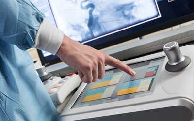
The Innova IGS 520 system offers a comprehensive suite of functionality customized for a wide range of EP procedures. With its 20.5 x 20.5 cm (8.1 in) square flat-panel digital detector, it offers excellent visualization of the heart and its chambers. The system is designed from the ground to provide the image clarity you need while helping you keep dose as low as possible. It includes features like Dose Personalization1, which gives you the tools to choose from up to four automatic exposure preferences for your system2. You can also modify3 any of these preferences in any clinical protocol to enable multi-procedure, multi-user customization and thus support well-informed decisions.
Innova IGS 520 features a 3.75 frames per second fluoroscopic acquisition mode optimized for EP procedures.


Power up your clinical decision making
The interventional field is growing with ever-expanding capabilities and migration to less invasive, safer and more cost-efficient procedures. With the new generation of GE's advanced interventional imaging software solutions, ASSIST, you can expand your clinical versatility and successfully plan, guide and assess increasingly sophisticated procedures with greater precision and dose efficiency.
EP catheter ablations
Cardiac ablation is an invasive procedure performed to correct heart arrhythmias such as atrial fibrillation. Ablation typically uses catheters - long, flexible tubes inserted through a vein in the groin and advanced to the heart to correct or scar the abnormal electrical circuits that are causing the arrhythmia. Ultimately, ablation prevents abnormal electrical signals from traveling through the heart and restores normal heart rhythm.


In clinical use, the results of the application of dose reduction techniques will vary depending on the clinical task, patient size, anatomical location and clinical practice. The Interventional radiologist, assisted by a physicist as necessary has to determine the appropriate settings for each specific clinical task.
1 – Option
2 – Except in following countries: Germany, Switzerland, Austria, New Zealand where that list is limited to 3 preferences
3 – By requesting it to your entity responsible for the servicing of your equipment
4 – Valve ASSIST 2 solution includes TAVI Analysis, HeartVision 2 and requires Volume Viewer, Volume Viewer International.
These applications are sold separately.
5 – Ultrasound imaging is not included in Valve ASSIST 2
6 – The U/S Image on the Large Display Monitor is for information. The primary display for U/S imaging is on the Ultrasound screen.
* Applications sold separately

The proprietary angiography flat-panel detector offers one of the industry’s highest ratings for Detective Quantum Efficiency (DQE), a parameter internationally acknowledged as an index of detector performance in contrast- and dose-limited imaging performed in clinical studies. High DQE enables better-quality images at the same dose, or the same quality image at a lower dose.

GE designs systems from the ground up with the tried and trusted GE imaging chain, optimized to provide the image clarity you need while helping you keep dose as low as possible. Our dose-reduction features empower you to easily optimize and personalize dose settings from the tableside, while maintaining clinical details you need to make well-informed decisions. But improving dose management takes a strategy – what we call the GE Blueprint. It includes low-dose imaging technologies for minimally invasive procedures, but also considers the people, culture and processes around them.